Collection Of Pus In The Pleural Cavity Quizlet
Breaking News Today
Mar 23, 2025 · 6 min read
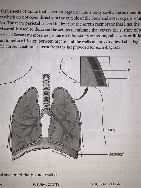
Table of Contents
Collection of Pus in the Pleural Cavity: A Comprehensive Guide
The presence of pus in the pleural cavity, a serious condition known as empyema, requires immediate medical attention. This condition represents a severe complication of various lung infections and other underlying diseases. Understanding the causes, symptoms, diagnosis, and treatment of empyema is crucial for effective management and improved patient outcomes. This comprehensive guide will delve deep into the topic, exploring its intricacies and providing a thorough understanding of this complex medical condition.
What is Empyema?
Empyema, also known as pyothorax, is defined as the accumulation of pus within the pleural space—the area between the lungs and the chest wall. This pus is typically composed of white blood cells, bacteria, and tissue debris, resulting from an infection. The pleural space, normally containing only a small amount of lubricating fluid, becomes inflamed and filled with this infectious material, compromising lung function and potentially leading to life-threatening complications.
Types of Empyema:
Empyema is broadly classified into several types based on its stage of development and the characteristics of the pus:
- Early-stage empyema (fibrinous): This is the initial stage, characterized by a fibrinous exudate—a thick, sticky fluid containing fibrin—within the pleural space. The pus is not yet fully formed.
- Localized empyema: The pus is confined to a specific area within the pleural space.
- Loculated empyema: The pus is enclosed within pockets or compartments within the pleural space, making drainage more challenging.
- Free-flowing empyema: The pus flows freely within the pleural space.
Causes of Empyema:
Empyema is typically a secondary infection, meaning it arises as a complication of another underlying condition. The most common causes include:
- Pneumonia: This is the leading cause, with bacterial pneumonia being the most frequent culprit. The infection can spread from the lung tissue into the pleural space.
- Lung abscess: A localized collection of pus within the lung tissue can rupture into the pleural space, causing empyema.
- Bronchiectasis: This condition, characterized by abnormal widening of the airways, can increase susceptibility to infection and subsequent empyema.
- Tuberculosis: Tuberculosis, a bacterial infection, can spread to the lungs and pleural space, leading to empyema.
- Trauma: Penetrating chest injuries can introduce bacteria into the pleural space, causing infection and empyema.
- Post-surgical complications: Following thoracic surgery, infection can occur, leading to the formation of empyema.
- Esophageal rupture: A tear in the esophagus can allow stomach contents to spill into the pleural space, causing infection.
- Cancer: Certain cancers, particularly lung cancer, can increase the risk of infection and empyema.
- Rheumatoid arthritis: This autoimmune disease can lead to pleural inflammation and an increased risk of infection.
- Immunodeficiency: Individuals with weakened immune systems are more susceptible to infections, including empyema.
Symptoms of Empyema:
The symptoms of empyema can vary depending on the severity and stage of the disease. Common symptoms include:
- Chest pain: Sharp, stabbing pain in the chest, often worsened by deep breaths or coughing.
- Shortness of breath (dyspnea): Difficulty breathing, often accompanied by rapid breathing (tachypnea).
- Cough: A persistent cough, which may produce purulent (pus-filled) sputum.
- Fever and chills: Signs of infection, often accompanied by sweating and general malaise.
- Fatigue: Extreme tiredness and weakness.
- Weight loss: Unexplained weight loss can occur in severe cases.
- Tachycardia: Rapid heart rate.
- Cyanosis: Bluish discoloration of the skin and mucous membranes due to decreased oxygen levels in the blood.
- Pleural effusion: The accumulation of fluid in the pleural space (this often precedes the development of empyema).
Diagnosis of Empyema:
Diagnosing empyema requires a combination of clinical evaluation and imaging studies:
- Physical examination: The physician will listen to the lungs using a stethoscope, looking for signs of reduced breath sounds or abnormal lung sounds.
- Chest X-ray: This is the primary imaging test used to detect pleural effusion and empyema. It reveals the presence of fluid or pus in the pleural space.
- Computed tomography (CT) scan: A CT scan provides a more detailed image of the chest, allowing for better visualization of the extent and location of the empyema. It can also help differentiate between different types of empyema, such as loculated or free-flowing empyema.
- Thoracentesis: A procedure where a needle is inserted into the pleural space to remove a sample of fluid for analysis. The fluid is examined for the presence of pus, bacteria, and white blood cells. This analysis helps confirm the diagnosis of empyema and identify the causative organism.
- Blood tests: Complete blood count (CBC) reveals the presence of infection (leukocytosis) and other relevant markers. Blood cultures may be taken to identify the infecting bacteria.
Treatment of Empyema:
The treatment of empyema aims to drain the pus from the pleural space and eliminate the infection. Treatment options include:
- Antibiotics: Antibiotic therapy is crucial in treating the underlying infection. The choice of antibiotic depends on the identification of the infecting organism through culture and sensitivity testing. Broad-spectrum antibiotics may be used initially while awaiting culture results.
- Chest tube drainage: This involves inserting a chest tube into the pleural space to drain the pus. This is the most common treatment for empyema. The tube is connected to a drainage system that removes the pus.
- Video-assisted thoracoscopic surgery (VATS): A minimally invasive surgical technique where a small incision is made to allow a camera and surgical instruments to access the pleural space. VATS allows for more effective drainage and removal of loculated empyema.
- Open thoracotomy: This is a more invasive surgical procedure, used in cases where other treatments have failed or in cases of very severe empyema. It involves making a larger incision in the chest wall to gain access to the pleural space.
- Fibrinolytic agents: These medications help break down the fibrinous material in the pleural space, aiding in drainage.
- Pleurodesis: A procedure performed after successful drainage to prevent recurrence. It involves the insertion of a chemical irritant into the pleural space to cause adhesion of the pleural layers, thus obliterating the pleural space.
Prognosis and Complications:
With prompt and appropriate treatment, the prognosis for empyema is generally good. However, delayed or inadequate treatment can lead to serious complications, including:
- Sepsis: A life-threatening condition caused by the body's overwhelming response to infection.
- Respiratory failure: Inability of the lungs to adequately oxygenate the blood.
- Lung abscess: Formation of a pus-filled cavity within the lung tissue.
- Bronchopleural fistula: An abnormal connection between the bronchi and the pleural space.
- Chronic empyema: Persistent infection and pus accumulation, despite treatment.
- Empyema necessitatis: A rare complication where the pus breaks through the chest wall, forming an external abscess.
Prevention of Empyema:
Preventing empyema focuses on preventing the underlying infections that can lead to this condition:
- Pneumonia vaccination: Vaccination can help prevent pneumonia, a major cause of empyema.
- Good hygiene practices: Frequent handwashing and avoiding close contact with infected individuals can help prevent respiratory infections.
- Prompt treatment of respiratory infections: Early and effective treatment of pneumonia and other respiratory infections can reduce the risk of developing empyema.
- Smoking cessation: Smoking significantly increases the risk of respiratory infections and should be avoided.
Conclusion:
Empyema is a serious condition requiring prompt diagnosis and treatment. Understanding the causes, symptoms, diagnosis, and treatment options is crucial for effective management and improved patient outcomes. Early intervention and adherence to the prescribed treatment plan significantly reduce the risk of complications and improve the chances of a full recovery. If you experience symptoms suggestive of empyema, seek immediate medical attention. Early diagnosis and appropriate treatment are essential for preventing serious complications and ensuring the best possible outcome. This detailed exploration of empyema should enhance understanding and aid in prompt medical intervention when necessary. Remember, always consult with a healthcare professional for any health concerns. This information is for educational purposes only and does not constitute medical advice.
Latest Posts
Latest Posts
-
Recycled Or Repurposed Munitions Are Considered Waste Military Munitions
Mar 25, 2025
-
Understanding The Benefits Of An Activity Can
Mar 25, 2025
-
Use Only A Bandsaw That Has A
Mar 25, 2025
-
As I Descend Go Down My Wet Suit Will
Mar 25, 2025
-
Which Of The Following Is A Structure Function Claim
Mar 25, 2025
Related Post
Thank you for visiting our website which covers about Collection Of Pus In The Pleural Cavity Quizlet . We hope the information provided has been useful to you. Feel free to contact us if you have any questions or need further assistance. See you next time and don't miss to bookmark.
