Correctly Label The Features Of The Larynx.
Breaking News Today
Mar 26, 2025 · 7 min read
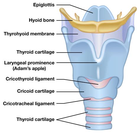
Table of Contents
Correctly Labeling the Features of the Larynx: A Comprehensive Guide
The larynx, often called the voice box, is a complex organ crucial for breathing, protecting the airway, and producing sound. Understanding its intricate anatomy is essential for various fields, including medicine, speech therapy, and singing. This comprehensive guide will delve into the detailed anatomy of the larynx, providing a clear and accurate understanding of its features and their functions. We'll explore the cartilages, muscles, membranes, ligaments, and spaces that constitute this vital organ, equipping you with the knowledge to correctly label its various components.
The Cartilages of the Larynx: The Foundation of Vocalization
The larynx is primarily composed of nine cartilages, connected by ligaments and membranes. These cartilages provide structural support and contribute to the complex mechanics of phonation (voice production). Let's examine each cartilage individually:
1. Thyroid Cartilage: The Largest and Most Prominent
The thyroid cartilage is the largest cartilage of the larynx, forming its anterior and lateral walls. Its shape resembles a shield, with two plates (alae) that meet anteriorly at the laryngeal prominence, commonly known as the "Adam's apple," which is more prominent in males due to hormonal influences during puberty. The superior border of the thyroid cartilage is attached to the hyoid bone via the thyrohyoid membrane, while the inferior border articulates with the cricoid cartilage. Its posterior horns (cornua) articulate with the cricoid cartilage, playing a crucial role in vocal fold adjustments.
2. Cricoid Cartilage: The Ring of Support
The cricoid cartilage sits inferior to the thyroid cartilage, forming the base of the larynx. Its shape resembles a signet ring, wider posteriorly than anteriorly. This unique shape is critical for its function in supporting the other laryngeal cartilages and providing an attachment point for various muscles and ligaments. The cricoid cartilage articulates with the first tracheal ring inferiorly and with the thyroid cartilage superiorly. Its crucial role in vocal fold adduction and abduction should not be overlooked.
3. Epiglottis: The Protective Lid
The epiglottis is a leaf-shaped cartilage located superior to the thyroid cartilage. Its primary function is to protect the airway during swallowing. When we swallow, the epiglottis folds down over the laryngeal inlet, preventing food and liquids from entering the trachea (windpipe). Its flexibility and positioning are critical for this protective mechanism. The epiglottis is attached to the hyoid bone and the thyroid cartilage by ligaments.
4. Arytenoid Cartilages: The Anchors of Vocal Fold Movement
The arytenoid cartilages are a pair of pyramid-shaped cartilages located on the superior posterior border of the cricoid cartilage. These cartilages are crucial for vocal fold movement. They possess vocal processes, which are attachment points for the vocal ligaments, and muscular processes, which serve as insertion points for intrinsic laryngeal muscles that control vocal fold abduction and adduction. The precise movements of the arytenoids are vital for phonation and breathing.
5. Corniculate Cartilages: Small but Significant
The corniculate cartilages are small, cone-shaped cartilages that articulate with the apexes of the arytenoid cartilages. They contribute to the structure of the aryepiglottic folds, which form part of the laryngeal inlet. While smaller, they still contribute to the overall mechanics of the larynx.
6. Cuneiform Cartilages: Embedded within the Aryepiglottic Folds
The cuneiform cartilages are small, rod-shaped cartilages embedded within the aryepiglottic folds. They provide structural support to these folds and are less prominent than other cartilages. However, their presence aids in shaping and supporting the upper portion of the larynx.
The Membranes and Ligaments: Connecting the Cartilages
The cartilages of the larynx are interconnected by several vital membranes and ligaments that provide stability, flexibility, and allow for the intricate movements necessary for phonation and respiration.
1. Thyrohyoid Membrane: Connecting Thyroid and Hyoid
The thyrohyoid membrane connects the superior border of the thyroid cartilage to the hyoid bone. It forms a strong connection that contributes to the stability of the larynx.
2. Cricotracheal Ligament: Linking Cricoid and Trachea
The cricotracheal ligament connects the inferior border of the cricoid cartilage to the first tracheal ring. This ligament provides structural support and continuity between the larynx and the trachea.
3. Cricothyroid Ligament: Crucial for Pitch Control
The cricothyroid ligament connects the cricoid cartilage to the thyroid cartilage. This ligament is vital for pitch control, as its stretching influences the tension of the vocal folds.
4. Quadrangular Membranes: Forming the Aryepiglottic Folds
The quadrangular membranes extend from the epiglottis to the arytenoid cartilages. Their free borders form the aryepiglottic folds, which contribute to the closure of the laryngeal inlet during swallowing.
5. Vocal Ligaments: The Foundation of Vocal Folds
The vocal ligaments are elastic ligaments that extend from the thyroid cartilage anteriorly to the vocal processes of the arytenoid cartilages posteriorly. These ligaments form the core of the vocal folds (vocal cords), which are responsible for sound production. Their tension and vibration are fundamental to voice quality.
The Muscles of the Larynx: The Power Behind Vocalization
The larynx contains both intrinsic and extrinsic muscles. Intrinsic muscles originate and insert within the larynx, controlling vocal fold movement and regulating laryngeal aperture. Extrinsic muscles connect the larynx to other structures, primarily the hyoid bone, and influence laryngeal position.
Intrinsic Laryngeal Muscles: Fine Motor Control of Phonation
The intrinsic laryngeal muscles are responsible for the fine motor control required for phonation. They include:
- Cricothyroid Muscle: This muscle tenses the vocal folds, increasing pitch.
- Thyroarytenoid Muscle: This muscle relaxes the vocal folds, decreasing pitch. It’s further divided into the vocalis and thyromuscularis muscles. The vocalis muscle is directly involved in vocal fold vibration.
- Posterior Cricoarytenoid Muscle: The only abductor of the vocal folds, opening the glottis for breathing.
- Lateral Cricoarytenoid Muscle: Adducts the vocal folds, bringing them together for phonation.
- Transverse Arytenoid Muscle: Adducts the arytenoid cartilages, contributing to vocal fold closure.
- Oblique Arytenoid Muscle: Adducts the arytenoid cartilages, also aiding in vocal fold closure.
Extrinsic Laryngeal Muscles: Positioning and Support
Extrinsic laryngeal muscles support and position the larynx within the neck. Key extrinsic muscles include:
- Sternothyroid Muscle: Depresses the larynx.
- Sternohyoid Muscle: Depresses the hyoid bone and indirectly influences laryngeal position.
- Thyrohyoid Muscle: Elevates the larynx.
- Digastric Muscle: Elevates the hyoid bone.
- Mylohyoid Muscle: Elevates the hyoid bone.
- Geniohyoid Muscle: Elevates the hyoid bone.
- Omohyoid Muscle: Depresses the hyoid bone.
Spaces and Cavities within the Larynx: Understanding the Functional Architecture
The larynx also encompasses several important spaces and cavities that contribute to its overall function:
- Laryngeal Ventricle: A space between the vocal folds and the vestibular folds (false vocal cords).
- Glottis: The space between the vocal folds, crucial for airflow during breathing and phonation. Its size and shape are dynamically regulated.
- Laryngeal Inlet: The superior opening of the larynx, guarded by the epiglottis.
- Subglottic Space: The area beneath the vocal folds, leading to the trachea.
Clinical Significance of Understanding Laryngeal Anatomy
A thorough understanding of laryngeal anatomy is crucial for various medical and therapeutic interventions. Accurate identification of laryngeal structures is paramount in diagnosing and treating conditions such as:
- Laryngeal Cancer: Early detection and accurate staging rely on a precise knowledge of laryngeal anatomy.
- Vocal Fold Nodules and Polyps: Treatment planning requires a detailed understanding of vocal fold structure and function.
- Laryngitis: Diagnosis and management of this inflammatory condition necessitates understanding the laryngeal structures involved.
- Tracheostomy: The placement of a tracheostomy tube requires accurate identification of anatomical landmarks.
- Laryngeal Trauma: Assessment and management of laryngeal injuries are facilitated by a thorough grasp of the anatomy.
Conclusion: Mastering the Laryngeal Landscape
This detailed exploration of the larynx highlights its intricate structure and the vital role each component plays in respiration, protection, and phonation. By accurately understanding and labeling the cartilages, membranes, ligaments, muscles, and spaces within the larynx, professionals in medicine, speech therapy, and related fields are better equipped to diagnose, treat, and manage a wide range of conditions impacting this critical organ. The information provided here serves as a comprehensive resource for mastering the complex anatomy of the larynx and fostering a deeper understanding of its significant function in the human body. Remember to consult reputable anatomical texts and resources for further learning and detailed visualization. Accurate labeling requires diligent study and practice, ultimately leading to a strong command of this complex and essential organ.
Latest Posts
Latest Posts
-
Ap Chem Unit 5 Progress Check Mcq
Mar 29, 2025
-
Which Of These Are True Of Tests For Online Courses
Mar 29, 2025
-
Drug Addiction Is A Clinical Diagnosis That Everfi
Mar 29, 2025
-
El Nacimiento Es El Fin De La Vida
Mar 29, 2025
-
A 26 Year Old Female Presents With Heavy Vaginal Bleeding
Mar 29, 2025
Related Post
Thank you for visiting our website which covers about Correctly Label The Features Of The Larynx. . We hope the information provided has been useful to you. Feel free to contact us if you have any questions or need further assistance. See you next time and don't miss to bookmark.
