Correctly Label The Following Anatomical Parts Of A Kidney.
Breaking News Today
Mar 14, 2025 · 7 min read
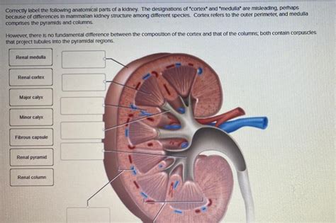
Table of Contents
Correctly Label the Following Anatomical Parts of a Kidney: A Comprehensive Guide
The kidney, a vital organ in the urinary system, plays a crucial role in maintaining overall health and homeostasis. Understanding its intricate anatomy is paramount for anyone studying biology, medicine, or simply fascinated by the human body. This comprehensive guide will delve deep into the anatomical structures of the kidney, providing detailed descriptions and visual aids to ensure a thorough understanding. We'll cover everything from the macroscopic structures visible to the naked eye to the microscopic components essential for its function.
Macroscopic Anatomy of the Kidney: An External Overview
Before diving into the microscopic intricacies, let's establish a solid understanding of the kidney's external features. Each kidney, roughly bean-shaped, is approximately 10-12 centimeters long, 5-7 centimeters wide, and 3-4 centimeters thick. Several key external features are readily identifiable:
1. Renal Capsule: The Protective Outer Layer
The renal capsule is a tough, fibrous membrane that encloses the entire kidney. This protective layer acts as a barrier, shielding the delicate renal tissue from external trauma and infection. Its smooth, glistening surface is readily visible during dissection.
2. Renal Hilum: The Gateway to the Kidney
The renal hilum, a concave medial border, is where the renal artery enters, the renal vein and ureter exit the kidney. This is the entry and exit point for the renal vasculature and the urinary outflow tract. Its location is crucial for understanding the blood supply and drainage of the kidney.
3. Renal Cortex: The Outer Region
The renal cortex is the reddish-brown outer region of the kidney, located just beneath the renal capsule. This region contains the majority of the nephrons, the functional units of the kidney responsible for filtering blood and producing urine. The cortex has a granular appearance due to the presence of numerous nephrons.
4. Renal Medulla: The Inner Region
Deep to the renal cortex lies the renal medulla, a darker, striated region. This area is organized into cone-shaped structures called renal pyramids. These pyramids are composed of parallel bundles of collecting ducts, which carry urine from the nephrons to the calyces. The striated appearance of the medulla is due to these tightly packed collecting ducts.
5. Renal Columns: Extensions of the Cortex
The renal columns, extensions of the renal cortex, extend inward between the renal pyramids. These columns provide structural support and facilitate the passage of blood vessels and nerves throughout the kidney. They contribute to the overall architecture of the renal medulla.
6. Renal Papillae: The Apices of the Pyramids
The apex of each renal pyramid is called the renal papilla. These papillae project into the minor calyces, small cup-like structures that collect urine produced by the nephrons. The papillae represent the point of urine drainage from the medulla.
7. Minor and Major Calyces: The Urine Collection System
The urine collected by the renal papillae flows into the minor calyces, which then merge to form larger structures known as major calyces. These major calyces ultimately converge to form the renal pelvis. The calyces are crucial in channeling urine from the pyramids to the renal pelvis.
8. Renal Pelvis: The Funnel-Shaped Reservoir
The renal pelvis, a funnel-shaped structure, acts as a reservoir for urine collected from the major calyces. It is located at the hilum and narrows to form the ureter, the tube that transports urine to the urinary bladder. The renal pelvis ensures a smooth flow of urine out of the kidney.
9. Ureter: The Pathway to the Bladder
The ureter is a muscular tube that extends from the renal pelvis to the urinary bladder. Its peristaltic contractions propel urine towards the bladder for storage and eventual elimination. The ureter's smooth muscle action is essential for urinary transport.
Microscopic Anatomy of the Kidney: The Nephron and its Components
The functional unit of the kidney is the nephron, a complex structure responsible for filtering blood and producing urine. Millions of nephrons are packed within the renal cortex and outer medulla. Each nephron consists of several key components:
1. Renal Corpuscle: The Filtration Unit
The renal corpuscle is the initial filtering unit of the nephron. It consists of two main structures:
- Glomerulus: A network of capillaries where blood filtration occurs. The high pressure within the glomerulus forces water and small solutes into Bowman's capsule.
- Bowman's Capsule: A double-walled cup-shaped structure surrounding the glomerulus. It receives the filtrate from the glomerulus, initiating the urine formation process. The capsule's specialized cells play a role in filtration.
2. Renal Tubule: The Processing Unit
The filtrate produced in the renal corpuscle then enters the renal tubule, a long, convoluted tube responsible for modifying the filtrate composition. The renal tubule is subdivided into several segments:
- Proximal Convoluted Tubule (PCT): The initial segment of the tubule, characterized by its extensive microvilli, enhances reabsorption of essential nutrients, water, and ions back into the bloodstream. This segment plays a critical role in conserving valuable substances.
- Loop of Henle: A U-shaped structure extending into the renal medulla. Its unique structure establishes an osmotic gradient crucial for concentrating urine. The descending and ascending limbs have distinct permeabilities, contributing to water reabsorption and ion transport.
- Distal Convoluted Tubule (DCT): The final segment of the nephron, responsible for fine-tuning the composition of the filtrate, particularly regarding electrolyte balance and acid-base homeostasis. Hormonal regulation plays a significant role in DCT function.
- Collecting Duct: While technically not part of the nephron itself, the collecting duct is where multiple distal convoluted tubules converge. It plays a crucial role in water reabsorption under the influence of antidiuretic hormone (ADH). This determines the final concentration of urine.
Blood Supply to the Kidney: A Vital Network
The kidney receives a rich blood supply through the renal artery, a branch of the abdominal aorta. This artery branches into progressively smaller arterioles, ultimately reaching the glomeruli, where blood filtration occurs. The filtered blood then exits the kidney through the renal vein, which drains into the inferior vena cava. This efficient blood supply ensures that waste products are efficiently removed from the blood and the body.
The renal artery branches into segmental arteries, then interlobar arteries that travel through the renal columns, then arcuate arteries which arch over the renal pyramids, and finally interlobular arteries that extend into the cortex. The blood then flows through the afferent arterioles into the glomeruli, and then exits through the efferent arterioles. This intricate network ensures a consistent and adequate blood supply for filtration.
Innervation of the Kidney: Neural Control
The kidneys receive sympathetic innervation from the renal plexus, a network of nerves originating from the celiac and superior mesenteric ganglia. This sympathetic innervation primarily regulates blood flow to the kidneys. It does not directly affect urine production. However, it plays a crucial role in adjusting renal blood flow in response to various physiological needs and stresses.
Clinical Significance: Understanding Kidney Diseases
Understanding the anatomy of the kidney is essential for diagnosing and treating various kidney diseases. Conditions like glomerulonephritis (inflammation of the glomeruli), kidney stones (formation of crystals in the urinary tract), and polycystic kidney disease (development of cysts in the kidneys) can significantly impair kidney function. A deep understanding of the kidney's structure and its delicate mechanisms is paramount in assessing and treating renal pathologies.
Furthermore, knowing the intricate pathways of urine flow from the renal papillae to the ureter is crucial in understanding the causes and progression of urinary tract infections and kidney stones. It provides a framework for visualizing how blockages occur, how infection spreads, and how treatment strategies are devised.
Conclusion: The Kidney – A Masterpiece of Engineering
The kidney, with its complex interplay of macroscopic and microscopic structures, is a remarkable organ. Its efficiency in filtering blood, regulating electrolyte balance, and maintaining overall homeostasis is a testament to the intricate design of the human body. This detailed exploration of its anatomy provides a solid foundation for further study and appreciation of this essential organ. The more we understand its structure, the better equipped we are to appreciate its vital role in maintaining health and well-being. By correctly labeling and understanding each component, we gain a deeper appreciation for the remarkable engineering of the human body and the significance of this often-overlooked organ.
Latest Posts
Latest Posts
-
After A Classified Document Is Leaked Online
Mar 14, 2025
-
Which Of The Following Represents Critical Information
Mar 14, 2025
-
Fundamentals Of Physics 12 Edition Instructors Solutions Manual Pdf
Mar 14, 2025
-
What Is Photosynthesis Check All That Apply
Mar 14, 2025
-
The Concept Overview Video Assignments Are Organized
Mar 14, 2025
Related Post
Thank you for visiting our website which covers about Correctly Label The Following Anatomical Parts Of A Kidney. . We hope the information provided has been useful to you. Feel free to contact us if you have any questions or need further assistance. See you next time and don't miss to bookmark.
