Correctly Label The Following Major Systemic Arteries.
Breaking News Today
Mar 13, 2025 · 6 min read
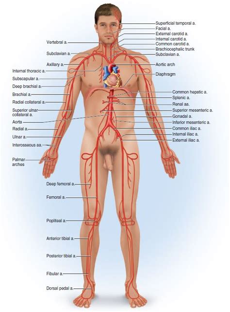
Table of Contents
Correctly Labeling the Major Systemic Arteries: A Comprehensive Guide
The systemic arteries form a complex network responsible for delivering oxygenated blood from the heart to the entire body. Understanding their precise labeling is crucial for medical professionals, students, and anyone interested in human anatomy and physiology. This comprehensive guide will delve into the major systemic arteries, providing detailed descriptions, branching patterns, and clinical significance. We will cover the arteries in a logical, hierarchical manner, starting from the heart and moving outwards to the periphery.
The Aorta: The Body's Primary Arterial Highway
The journey begins with the aorta, the largest artery in the body. Originating from the left ventricle of the heart, it’s divided into several segments:
1. Ascending Aorta:
The ascending aorta is the initial portion, arising directly from the left ventricle. It's relatively short, giving rise to two crucial branches:
- Right Coronary Artery: Supplies blood to the right atrium, right ventricle, and parts of the left ventricle. Blockages here can lead to significant cardiac issues.
- Left Coronary Artery: Branches into the circumflex artery and the anterior interventricular artery, supplying blood to the majority of the left ventricle and parts of the right ventricle. Occlusion in this artery is a leading cause of heart attacks.
2. Aortic Arch:
The aortic arch curves posteriorly and to the left, giving rise to three major branches:
- Brachiocephalic Trunk: This is the first branch, quickly dividing into the right common carotid artery and the right subclavian artery.
- Right Common Carotid Artery: Supplies blood to the right side of the head and neck. It further bifurcates into the internal and external carotid arteries.
- Right Subclavian Artery: Supplies blood to the right upper limb and parts of the neck and thorax.
- Left Common Carotid Artery: Supplies blood to the left side of the head and neck, mirroring the right common carotid in its branching pattern.
- Left Subclavian Artery: Supplies blood to the left upper limb, mirroring the right subclavian artery.
3. Thoracic Aorta:
Descending from the aortic arch, the thoracic aorta travels through the thoracic cavity, giving rise to numerous smaller branches supplying the thoracic organs and the intercostal muscles:
- Visceral Branches: These supply blood to the lungs (bronchial arteries), esophagus (esophageal arteries), and pericardium (pericardial arteries).
- Parietal Branches: These include the posterior intercostal arteries, supplying the muscles and tissues of the thoracic wall. These arteries are crucial for the oxygenation of the back muscles and ribcage.
4. Abdominal Aorta:
Continuing below the diaphragm, the abdominal aorta is the largest segment, providing blood supply to the abdominal organs and lower limbs. It's characterized by its numerous branches:
- Celiac Trunk: A major branch arising near the level of the first lumbar vertebra, it further divides into three arteries:
- Left Gastric Artery: Supplies the stomach and lesser curvature.
- Splenic Artery: Supplies the spleen, pancreas, and stomach.
- Common Hepatic Artery: Supplies the liver, gallbladder, and stomach. This artery is often a target for angiographic procedures.
- Superior Mesenteric Artery: Supplies blood to most of the small intestine and part of the large intestine (ascending colon and transverse colon). Its integrity is crucial for proper digestion and nutrient absorption.
- Inferior Mesenteric Artery: Supplies blood to the distal parts of the large intestine (descending colon, sigmoid colon, and rectum). Problems here can manifest as bowel ischemia.
- Renal Arteries: Two branches, one for each kidney, vital for renal function and blood filtration. Kidney disease can significantly impact renal artery health.
- Gonadal Arteries: These supply the gonads (testes in males and ovaries in females). Their function is essential for reproductive health.
- Lumbar Arteries: Supply the lumbar muscles and the posterior abdominal wall. These arteries are often involved in back pain syndromes.
- Common Iliac Arteries: These are the terminal branches of the abdominal aorta, each dividing into the internal and external iliac arteries.
- Internal Iliac Arteries: Supply blood to the pelvic organs and gluteal region.
- External Iliac Arteries: Continue into the lower limbs as the femoral arteries.
Arteries of the Lower Limbs:
The external iliac artery continues into the thigh as the femoral artery. This is a critical artery, often used for arterial access in medical procedures. The femoral artery gives rise to several branches before becoming the popliteal artery behind the knee. The popliteal artery further branches into the anterior and posterior tibial arteries and the fibular artery, supplying blood to the lower leg and foot.
- Femoral Artery: This major artery supplies blood to the thigh and its various muscles and tissues. Injury to this vessel can lead to severe blood loss.
- Popliteal Artery: Located behind the knee joint, this artery is crucial for supplying blood to the muscles of the lower leg.
- Anterior Tibial Artery: Supplies blood to the anterior compartment of the leg and dorsum of the foot.
- Posterior Tibial Artery: Supplies blood to the posterior compartment of the leg and plantar surface of the foot.
- Fibular Artery: Supplies blood to the lateral compartment of the leg.
Arteries of the Upper Limbs:
The subclavian artery continues into the arm as the axillary artery, which then becomes the brachial artery. The brachial artery bifurcates into the radial and ulnar arteries at the elbow. These arteries and their branches supply the muscles, bones, and tissues of the arm and hand.
- Axillary Artery: This artery runs through the armpit and supplies blood to the shoulder and chest muscles.
- Brachial Artery: This artery runs down the arm and supplies blood to the biceps and triceps muscles. The brachial pulse is frequently checked during medical examinations.
- Radial Artery: Located on the thumb side of the forearm, this artery is easily palpable and is frequently used to check the pulse.
- Ulnar Artery: Located on the little finger side of the forearm, this artery runs alongside the ulna bone.
Clinical Significance and Diagnostic Imaging:
Understanding the systemic arteries is essential for diagnosing and treating various vascular diseases. Conditions such as atherosclerosis (plaque buildup in the arteries), aneurysms (bulges in the arterial walls), and embolisms (blockages in the arteries) can have devastating consequences if left untreated.
Diagnostic imaging techniques, such as angiography, computed tomography angiography (CTA), and magnetic resonance angiography (MRA), are invaluable in visualizing the arteries and detecting abnormalities. These techniques enable medical professionals to precisely identify the location and extent of vascular diseases and guide treatment decisions.
Conclusion:
This comprehensive guide provides a detailed overview of the major systemic arteries. Precise labeling and understanding of these arteries are crucial for comprehending blood flow dynamics, diagnosing vascular diseases, and providing appropriate medical interventions. Further exploration of individual arterial branches and their detailed anatomical relationships will enhance your understanding of this complex and vital system. Remember to consult reputable anatomical resources and textbooks for a more in-depth study of this fascinating subject. The accurate labeling of these arteries is paramount for effective medical practice and patient care. Continuous learning and review are key to maintaining proficiency in this area. The detailed understanding of the systemic arterial system is a cornerstone of medical knowledge.
Latest Posts
Latest Posts
-
A 30 Year Old Woman With A History Of Alcoholism
Mar 13, 2025
-
Which Groups Best Fit The Theistic Worldview
Mar 13, 2025
-
You Tap And Shout To Check For Responsiveness
Mar 13, 2025
-
Which Is Not A Function Of Bone
Mar 13, 2025
-
Swapping Items Between Memory And Storage Is Called
Mar 13, 2025
Related Post
Thank you for visiting our website which covers about Correctly Label The Following Major Systemic Arteries. . We hope the information provided has been useful to you. Feel free to contact us if you have any questions or need further assistance. See you next time and don't miss to bookmark.
