Correctly Label The Following Parts Of A Renal Corpuscle.
Breaking News Today
Mar 14, 2025 · 6 min read
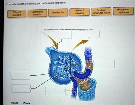
Table of Contents
Correctly Label the Following Parts of a Renal Corpuscle: A Deep Dive into Nephron Structure and Function
The renal corpuscle, the initial filtering unit of the nephron, plays a pivotal role in the intricate process of urine formation. Understanding its components is crucial to grasping the complexities of renal physiology. This comprehensive guide will delve into the detailed structure and function of the renal corpuscle, enabling you to correctly label its key parts with confidence. We'll explore the glomerulus, Bowman's capsule, and the crucial interplay between them, highlighting their individual contributions to filtration and the overall health of the urinary system.
The Glomerulus: A Network of Filtration
The glomerulus, a tuft of capillaries, forms the heart of the renal corpuscle. This isn't your average capillary bed; the glomerular capillaries are fenestrated, meaning they possess numerous pores or windows. These fenestrations allow for the passage of water and small solutes while largely excluding larger molecules like proteins and blood cells. This selective permeability is the first crucial step in blood filtration.
Key Features of the Glomerular Capillaries:
-
Fenestrations: These pores are approximately 70-100 nm in diameter, preventing the passage of larger molecules while allowing smaller ones to pass.
-
Glomerular Basement Membrane (GBM): This specialized extracellular matrix lies between the endothelial cells of the glomerular capillaries and the podocytes of Bowman's capsule. The GBM acts as a further filtration barrier, restricting the passage of larger molecules and negatively charged proteins based on size and charge selectivity. Its intricate composition includes type IV collagen, laminin, and proteoglycans, contributing to its filtering capabilities. Damage to the GBM, as seen in certain glomerulonephritides, can significantly impair filtration.
-
Podocytes: These highly specialized epithelial cells cling to the outer surface of the glomerular capillaries. Their unique structure, featuring foot-like processes called pedicels, interdigitates to form filtration slits. These slits, spanned by a specialized membrane called the slit diaphragm, represent the final barrier to filtration, further refining the selectivity process and restricting the passage of even smaller proteins and macromolecules. The slit diaphragm is particularly effective at preventing the passage of albumin, a crucial protein that should remain in the bloodstream. Dysfunction of podocytes is a major factor in various kidney diseases.
Bowman's Capsule: Encapsulating the Filtration Process
Bowman's capsule, a double-walled epithelial cup, surrounds the glomerulus. It's composed of two layers: the parietal layer and the visceral layer.
Understanding the Layers of Bowman's Capsule:
-
Parietal Layer: This outer layer is a simple squamous epithelium. It forms the structural framework of the capsule and acts as a passive supportive structure. It does not directly participate in the filtration process.
-
Visceral Layer: This inner layer is where the action is. The visceral layer is made up of the aforementioned podocytes, which are intimately associated with the glomerular capillaries. It's the interaction between the podocytes and the glomerular basement membrane that determines what passes through into the capsular space.
The Capsular Space: Where Filtrate Begins
The space between the parietal and visceral layers is the capsular space, also known as Bowman's space. This is where the filtered fluid, now called glomerular filtrate, accumulates. The filtrate is essentially a protein-free plasma that now begins its journey through the nephron towards the final formation of urine.
The Filtration Process: A Precisely Orchestrated Event
The filtration process within the renal corpuscle is a remarkable feat of biological engineering. It's driven by the glomerular filtration pressure (GFP), a complex interplay of forces that favor filtration:
-
Glomerular capillary blood pressure: This force pushes fluid out of the capillaries and into Bowman's space. It is the primary driving force behind filtration.
-
Capsular hydrostatic pressure: This pressure opposes filtration by pushing fluid back into the glomerular capillaries.
-
Blood colloid osmotic pressure: This pressure, caused by the presence of proteins in the blood, also opposes filtration by drawing fluid back into the capillaries.
The GFP is the net result of these forces; a carefully balanced equation that determines the rate of filtration. Any alteration in these pressures, such as increased blood pressure or decreased protein levels, will directly impact the filtration rate.
The Juxtaglomerular Apparatus: Regulation and Feedback
The renal corpuscle doesn't operate in isolation. It's closely integrated with the juxtaglomerular apparatus (JGA), a specialized structure located at the junction between the distal convoluted tubule and the afferent arteriole. The JGA plays a critical role in regulating glomerular filtration rate (GFR) through a sophisticated feedback mechanism. Components of the JGA include:
-
Juxtaglomerular cells: These specialized smooth muscle cells in the afferent arteriole secrete renin, a hormone that plays a crucial role in blood pressure regulation and sodium balance. Renin activates the renin-angiotensin-aldosterone system (RAAS), a powerful hormonal pathway that affects GFR and overall blood pressure.
-
Macula densa: These specialized cells in the distal convoluted tubule detect changes in the sodium concentration of the filtrate. They signal the juxtaglomerular cells to adjust renin secretion in response to changes in GFR and sodium levels.
The JGA's intricate interplay ensures that GFR remains relatively stable despite fluctuations in blood pressure and other physiological factors.
Clinical Significance: Diseases Affecting the Renal Corpuscle
Dysfunction of the renal corpuscle can lead to various kidney diseases. Conditions such as glomerulonephritis, characterized by inflammation of the glomeruli, can significantly impair filtration, leading to proteinuria (protein in the urine) and hematuria (blood in the urine). Similarly, damage to podocytes can result in nephrotic syndrome, marked by significant protein loss in the urine. Understanding the structure and function of the renal corpuscle is crucial for diagnosing and managing these conditions. Early detection and appropriate intervention are essential to preserving renal function.
Labeling the Renal Corpuscle: A Practical Exercise
Now that we've thoroughly explored the structure and function of the renal corpuscle, let's summarize the key components to effectively label a diagram:
- Glomerulus: The network of fenestrated capillaries.
- Bowman's Capsule: The double-walled epithelial cup.
- Parietal Layer of Bowman's Capsule: The outer, simple squamous epithelium.
- Visceral Layer of Bowman's Capsule: The inner layer containing podocytes.
- Podocytes: Specialized epithelial cells with foot-like processes (pedicels).
- Pedicels: Interdigitating foot-like processes of podocytes.
- Filtration Slits: Gaps between pedicels.
- Slit Diaphragm: Specialized membrane spanning the filtration slits.
- Glomerular Basement Membrane (GBM): The specialized extracellular matrix between the glomerular capillaries and podocytes.
- Capsular Space (Bowman's Space): The space between the parietal and visceral layers, where filtrate collects.
- Afferent Arteriole: The vessel supplying blood to the glomerulus.
- Efferent Arteriole: The vessel draining blood from the glomerulus.
- Juxtaglomerular Apparatus (JGA): (Located at the junction of the distal convoluted tubule and afferent arteriole – though not strictly part of the renal corpuscle, it’s functionally linked). Includes juxtaglomerular cells and macula densa.
By understanding these components and their intricate interactions, you can accurately label the parts of a renal corpuscle and gain a deeper appreciation for the vital role it plays in maintaining overall health and homeostasis. Remember, the precise filtration process carried out by the renal corpuscle is fundamental to the urinary system's function in excreting waste products and regulating fluid and electrolyte balance. Any disruption to this delicate balance can have far-reaching consequences. Continued study and comprehension of this complex system are essential for anyone pursuing knowledge in the fields of anatomy, physiology, and medicine.
Latest Posts
Latest Posts
-
What Would Be An Expense Factor In An Insurance Program
May 09, 2025
-
The First Space Zone Is Directly Above The Vehicle
May 09, 2025
-
Moviegoers Burst Into Laughter When A Black Leather Clad
May 09, 2025
-
Choose The Best Lewis Structure For Ocl2
May 09, 2025
-
Which Of The Following Statements About Osmosis Is False
May 09, 2025
Related Post
Thank you for visiting our website which covers about Correctly Label The Following Parts Of A Renal Corpuscle. . We hope the information provided has been useful to you. Feel free to contact us if you have any questions or need further assistance. See you next time and don't miss to bookmark.