Correctly Label The Intrinsic Muscles Of The Hand
Breaking News Today
Apr 03, 2025 · 6 min read

Table of Contents
Correctly Labeling the Intrinsic Muscles of the Hand: A Comprehensive Guide
The intrinsic muscles of the hand are a complex group of muscles responsible for the intricate movements of the fingers and thumb. Understanding their anatomy and function is crucial for healthcare professionals, artists, musicians, and anyone interested in the intricacies of human anatomy. This comprehensive guide provides a detailed overview, aiming to help you correctly label and understand these essential muscles. We'll cover their origins, insertions, actions, innervation, and clinical relevance, ensuring a thorough and accessible understanding.
Understanding the Intrinsic Hand Muscles: A Layered Approach
The intrinsic muscles of the hand are located entirely within the hand itself, unlike extrinsic muscles that originate in the forearm. They are responsible for fine motor control, allowing for precise movements needed for tasks like writing, playing musical instruments, and manipulating small objects. These muscles are broadly categorized into three groups: thenar, hypothenar, and midpalmar muscles. Let's explore each group in detail:
I. Thenar Muscles: The Thumb's Powerhouse
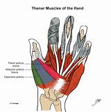
The thenar muscles are situated at the base of the thumb, enabling its unique range of motion, crucial for opposition (touching the thumb to other fingers) and precision grips.
1. Abductor pollicis brevis:
- Origin: Scapholunate ligament and flexor retinaculum.
- Insertion: Radial side of the proximal phalanx of the thumb.
- Action: Abducts the thumb. Think of it as pulling the thumb away from the index finger.
- Innervation: Median nerve (recurrent branch).
2. Flexor pollicis brevis:
- Origin: Flexor retinaculum and the trapezium and scaphoid bones.
- Insertion: Radial side of the proximal phalanx of the thumb.
- Action: Flexes the thumb at the metacarpophalangeal joint. This brings the thumb towards the palm.
- Innervation: Median nerve (recurrent branch). Superficial head is also innervated by the ulnar nerve.
3. Opponens pollicis:
- Origin: Trapezium and flexor retinaculum.
- Insertion: Entire length of the radial metacarpal bone.
- Action: Opposes the thumb, bringing the thumb across the palm to touch the other fingers. Essential for grasping objects.
- Innervation: Median nerve (recurrent branch).
4. Adductor pollicis:
- Origin: Two heads: oblique head (capitate and bases of the second and third metacarpals) and transverse head (bases of the second and third metacarpals).
- Insertion: Ulnar side of the base of the proximal phalanx of the thumb.
- Action: Adducts the thumb, moving it towards the index finger. Plays a critical role in power grips.
- Innervation: Ulnar nerve (deep branch). This is a key differentiator from the other thenar muscles.
II. Hypothenar Muscles: The Little Finger's Support System
The hypothenar muscles, located on the ulnar side of the hand at the base of the little finger, are responsible for its movement and stability.
1. Abductor digiti minimi:
- Origin: Pisiform bone and flexor retinaculum.
- Insertion: Ulnar side of the proximal phalanx of the little finger.
- Action: Abducts the little finger, pulling it away from the ring finger.
- Innervation: Ulnar nerve.
2. Flexor digiti minimi brevis:
- Origin: Hamate and flexor retinaculum.
- Insertion: Ulnar side of the proximal phalanx of the little finger.
- Action: Flexes the little finger at the metacarpophalangeal joint.
- Innervation: Ulnar nerve.
3. Opponens digiti minimi:
- Origin: Hamate and flexor retinaculum.
- Insertion: Ulnar side of the fifth metacarpal bone.
- Action: Opposes the little finger, slightly pulling it across the palm towards the thumb. Though less pronounced than thumb opposition.
- Innervation: Ulnar nerve.
III. Midpalmar Muscles: The Interosseous and Lumbricals
These muscles are centrally located within the hand, crucial for finger flexion, extension, abduction, and adduction. They are vital for precise and powerful hand movements.
1. Lumbricals (4 muscles):
- Origin: Tendons of the flexor digitorum profundus.
- Insertion: Extensor expansion of the index, middle, ring, and little fingers.
- Action: Flex the metacarpophalangeal joints and extend the interphalangeal joints of the fingers 2-5. They are often described as contributing to both flexion and extension depending on the joint in question.
- Innervation: Lumbricals 1 and 2 are innervated by the median nerve, while lumbricals 3 and 4 are innervated by the ulnar nerve.
2. Dorsal Interossei (4 muscles):
- Origin: Adjacent metacarpal bones.
- Insertion: Proximal phalanx and extensor expansion of the index, middle, ring, and little fingers.
- Action: Abduct the fingers (away from the middle finger). They also assist in flexing the metacarpophalangeal joints and extending the interphalangeal joints.
- Innervation: Ulnar nerve.
3. Palmar Interossei (3 muscles):
- Origin: Adjacent metacarpal bones.
- Insertion: Proximal phalanx and extensor expansion of the index, ring, and little fingers.
- Action: Adduct the fingers (towards the middle finger). They also assist in flexing the metacarpophalangeal joints and extending the interphalangeal joints.
- Innervation: Ulnar nerve.
Clinical Relevance: Conditions Affecting Intrinsic Hand Muscles
Understanding the intrinsic muscles is crucial in diagnosing and treating various hand conditions. Injuries and diseases can significantly impact their function, leading to decreased hand dexterity and grip strength. Some clinical conditions impacting these muscles include:
- Carpal Tunnel Syndrome: Compression of the median nerve at the wrist can affect the thenar muscles, causing weakness and atrophy.
- Ulnar Nerve Entrapment: Compression of the ulnar nerve can lead to weakness or paralysis of the hypothenar muscles, interossei, and the medial lumbricals. This can result in a condition known as "claw hand."
- Dupuytren's Contracture: A thickening and shortening of the palmar fascia can cause contractures of the fingers, particularly affecting the ring and little fingers, indirectly impacting the function of the intrinsic muscles.
- Hand Fractures: Fractures of the metacarpals can damage the attachments of the intrinsic muscles, leading to functional impairments.
- Muscle Strains and Sprains: Overuse or injury can lead to strains or sprains of the intrinsic muscles, resulting in pain, weakness, and decreased range of motion.
Memorization Techniques and Practical Application
Learning the intrinsic muscles can be challenging due to their number and intricate arrangement. Here are some helpful techniques for memorization and practical application:
- Mnemonic Devices: Create acronyms or rhymes to remember the origins, insertions, and actions of each muscle. For example, you could create a mnemonic for the thenar muscles based on their actions: Abducts, Flexes, Opposes, Adducts (AFOA).
- Visual Aids: Use anatomical diagrams, models, and interactive resources to visualize the muscles' positions and relationships.
- Palpation: Practice palpating the muscles on a willing participant to gain a better understanding of their location and texture. This tactile learning method can significantly aid memorization.
- Clinical Correlation: Relate the muscles' functions to common hand movements and clinical conditions. This contextual learning helps solidify your understanding.
- Repeated Practice: Consistent review and self-testing are crucial for long-term retention of anatomical information. Use flashcards, quizzes, and practice labeling diagrams.
Advanced Considerations and Further Exploration
Beyond the basics, further exploration of the intrinsic hand muscles can include:
- Muscle Synergies: Investigate how multiple intrinsic muscles work together to perform complex movements.
- Biomechanics: Analyze the forces and levers involved in hand movements, considering the roles of individual muscles.
- Comparative Anatomy: Compare the hand muscles of different species to understand evolutionary adaptations.
- Surgical Anatomy: Explore the surgical approaches and considerations related to the intrinsic hand muscles.
By mastering the labeling and understanding of the intrinsic hand muscles, you gain a deeper appreciation for the complexity and elegance of human anatomy. This knowledge is invaluable for professionals and enthusiasts alike, providing a solid foundation for further learning and applications in various fields. Remember to utilize the memorization techniques and continually review the information for optimal retention and understanding. This detailed guide serves as a starting point for a deeper dive into this fascinating and functional aspect of human anatomy. Through persistent study and practical application, you can achieve a comprehensive grasp of these vital muscles and their roles in hand function.
Latest Posts
Latest Posts
-
Degree Of Loudness Or Softness In Music
Apr 04, 2025
-
Select The Appropriate Term For Its Respective Definition
Apr 04, 2025
-
Strategic Intent Refers To A Situation Where A Company
Apr 04, 2025
-
The Reasons That Nations Trade Includes The Fact That
Apr 04, 2025
-
The First Sphincter Encountered In The Alimentary Canal Is The
Apr 04, 2025
Related Post
Thank you for visiting our website which covers about Correctly Label The Intrinsic Muscles Of The Hand . We hope the information provided has been useful to you. Feel free to contact us if you have any questions or need further assistance. See you next time and don't miss to bookmark.
