During Your Assessment Of A Patient With Blunt Chest Trauma
Breaking News Today
Mar 21, 2025 · 6 min read
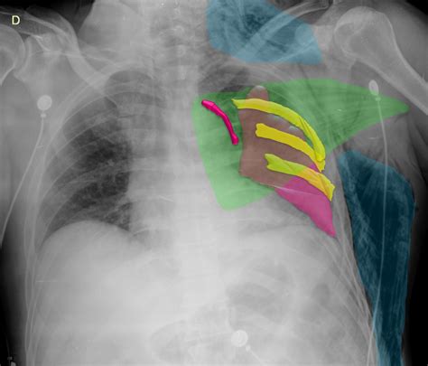
Table of Contents
During Your Assessment of a Patient with Blunt Chest Trauma
Blunt chest trauma (BCT) represents a significant clinical challenge, encompassing a wide spectrum of injuries ranging from minor contusions to life-threatening conditions. A thorough and systematic assessment is crucial for timely diagnosis and management, ultimately impacting patient outcomes. This article delves into the comprehensive evaluation of a patient presenting with BCT, covering the initial approach, detailed physical examination, investigative procedures, and considerations for specific injuries.
The Initial Approach: ABCDEs and Beyond
The initial management of any trauma patient, including those with BCT, follows the established ABCDE approach:
A - Airway: Maintaining a patent airway is paramount. Assess for airway compromise due to factors like maxillofacial injuries, cervical spine injury, or the presence of blood or secretions. Consider the need for advanced airway management, such as endotracheal intubation, if necessary. Always remember to protect the cervical spine during airway manipulation.
B - Breathing: Evaluate respiratory effort, rate, and depth. Auscultate the lungs for breath sounds, noting the presence of diminished breath sounds, wheezes, or rales. Observe the chest wall for paradoxical movement (flail chest), crepitus (subcutaneous emphysema), or tracheal deviation. Assess oxygen saturation (SpO2) using pulse oximetry. Supplemental oxygen should be administered immediately.
C - Circulation: Assess heart rate, blood pressure, and capillary refill time. Look for signs of shock, including tachycardia, hypotension, and pallor. Establish intravenous (IV) access, and consider fluid resuscitation if indicated. Monitor for signs of internal bleeding, such as distended neck veins (suggestive of pericardial tamponade) or a progressively expanding hematoma.
D - Disability: Briefly assess neurological status using the Glasgow Coma Scale (GCS). Look for signs of head injury, such as altered mental status, pupillary abnormalities, or focal neurological deficits.
E - Exposure: Completely undress the patient to identify all injuries. Maintain patient warmth to prevent hypothermia.
Detailed Physical Examination of the Chest
Following the initial ABCDE assessment, a detailed physical examination of the chest is essential:
1. Inspection:
- Chest Wall Deformities: Look for flail chest (paradoxical movement of a segment of the chest wall), rib fractures, sternal fractures, or penetrating wounds.
- Respiratory Effort: Observe the respiratory rate, depth, and pattern. Assess for use of accessory muscles, indicating respiratory distress.
- Skin: Check for abrasions, contusions, lacerations, or subcutaneous emphysema (air trapped beneath the skin). Subcutaneous emphysema suggests pneumothorax or tracheobronchial injury.
- Tracheal Deviation: Observe for tracheal deviation, which may indicate a tension pneumothorax.
2. Palpation:
- Chest Wall Tenderness: Palpate the chest wall for tenderness, identifying areas of rib fractures or other injuries.
- Crepitus: Palpate for crepitus, a crackling sensation indicating the presence of subcutaneous air.
- Heart Sounds: Palpate the precordium for thrills or heaves.
3. Auscultation:
- Breath Sounds: Auscultate lung fields systematically, comparing both sides. Listen for diminished breath sounds, wheezes, rales, or the absence of breath sounds. Diminished or absent breath sounds suggest pneumothorax or hemothorax.
- Heart Sounds: Auscultate heart sounds for murmurs, rubs, or other abnormalities. A pericardial friction rub may indicate pericarditis.
Investigative Procedures
Following the physical examination, various diagnostic tests may be indicated:
- Chest X-ray: A crucial initial imaging modality. It helps identify pneumothorax, hemothorax, rib fractures, flail chest, and other intrathoracic injuries. It can also assess for widening of the mediastinum, suggesting aortic injury.
- Computed Tomography (CT) Scan: CT scan provides a more detailed assessment of intrathoracic injuries, including lung contusions, rib fractures, vascular injuries, and cardiac injuries.
- Ultrasound: Focused Assessment with Sonography for Trauma (FAST) exam can rapidly assess for free fluid in the pericardial sac (pericardial effusion), pleural space (hemothorax or pneumothorax), or peritoneal cavity.
- Electrocardiogram (ECG): ECG helps detect cardiac contusion, arrhythmias, and other cardiac complications.
- Arterial Blood Gases (ABGs): ABGs provide information about oxygenation and ventilation.
- Laboratory Tests: Complete blood count (CBC) to assess for anemia or infection. Coagulation studies to assess for bleeding disorders.
Specific Injuries and Considerations
Several specific chest injuries are frequently associated with blunt chest trauma:
1. Pneumothorax:
A pneumothorax is the presence of air in the pleural space, causing lung collapse. Tension pneumothorax, a life-threatening complication, occurs when air enters the pleural space but cannot escape, leading to progressive lung collapse and cardiovascular compromise. Treatment involves needle decompression followed by chest tube insertion.
2. Hemothorax:
Hemothorax is the accumulation of blood in the pleural space. It can be caused by injury to the lung, intercostal vessels, or other intrathoracic vessels. Treatment typically involves chest tube insertion and drainage.
3. Flail Chest:
A flail chest occurs when multiple adjacent ribs are fractured in two or more places, creating a segment of the chest wall that moves paradoxically during breathing. This paradoxical movement impairs ventilation and can lead to respiratory distress. Management may involve pain control, assisted ventilation, and occasionally surgical stabilization.
4. Cardiac Contusion:
Cardiac contusion is bruising of the heart muscle. It can result in arrhythmias, heart failure, or other cardiac complications. ECG monitoring and cardiac enzyme levels are important for assessment.
5. Aortic Injury:
Aortic injury is a life-threatening complication that can result from high-energy blunt chest trauma. It can lead to rapid exsanguination and death. Diagnosis often involves CT angiography.
6. Tracheobronchial Injury:
Tracheobronchial injuries are relatively rare but potentially life-threatening. They can cause respiratory compromise and require urgent surgical intervention.
7. Esophageal Injury:
Esophageal injury is another less common but serious complication. It often requires surgical repair.
Ongoing Monitoring and Management
Following initial assessment and treatment, ongoing monitoring is crucial. This includes:
- Respiratory Monitoring: Closely monitor respiratory rate, depth, and oxygen saturation. Assess for signs of respiratory distress.
- Hemodynamic Monitoring: Monitor blood pressure, heart rate, and urine output.
- Cardiac Monitoring: Continuous ECG monitoring is recommended, especially in patients with cardiac contusion.
- Pain Management: Adequate pain control is essential to facilitate breathing and improve patient comfort.
- Serial Chest X-rays: Serial chest X-rays are helpful to monitor the effectiveness of treatment for pneumothorax or hemothorax.
Predicting Outcomes and Prognosis
The prognosis for patients with BCT varies greatly depending on the severity of the injuries. Factors that influence prognosis include the mechanism of injury, the presence of associated injuries, the effectiveness of initial resuscitation, and the patient's overall health. Early diagnosis and appropriate management are crucial for improving patient outcomes.
Keywords: blunt chest trauma, chest trauma, trauma assessment, ABCDE, pneumothorax, hemothorax, flail chest, cardiac contusion, aortic injury, chest x-ray, CT scan, ultrasound, management, treatment, prognosis
This article provides a comprehensive overview of the assessment and management of patients with blunt chest trauma. It's crucial to remember that this information is for educational purposes only and should not be considered medical advice. Always consult with qualified medical professionals for diagnosis and treatment of any medical condition. The management of blunt chest trauma requires a multidisciplinary approach involving emergency medical services, trauma surgeons, intensivists, and other specialists. Continuous professional development and adherence to established guidelines are essential for optimal patient care.
Latest Posts
Latest Posts
-
Which Of The Following Statements Are True Of Teams
Mar 27, 2025
-
A Group Of Biologists Is Studying The Competitive Relationships
Mar 27, 2025
-
The Keyword Tyranny In This Poster Is Primarily Used To
Mar 27, 2025
-
Irene Todavia No 1 Of 2 Lista Para Salir
Mar 27, 2025
-
A Bird Building Their Nest In A Tree
Mar 27, 2025
Related Post
Thank you for visiting our website which covers about During Your Assessment Of A Patient With Blunt Chest Trauma . We hope the information provided has been useful to you. Feel free to contact us if you have any questions or need further assistance. See you next time and don't miss to bookmark.
