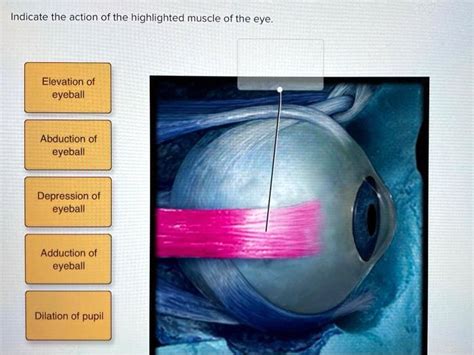
Indicate Which Muscle Is Highlighted In The Image.
Breaking News Today
Apr 02, 2025 · 5 min read

Table of Contents
Identifying Highlighted Muscles: A Comprehensive Guide
Identifying muscles in images requires a keen understanding of human anatomy. This article will serve as a comprehensive guide to identifying highlighted muscles, offering tips and tricks for accurate identification, regardless of the image's quality or angle. We will cover various approaches, from utilizing anatomical landmarks to understanding muscle actions and origins/insertions. We'll also explore common pitfalls and offer resources for further learning.
Importance of Accurate Muscle Identification
Accurate muscle identification is crucial in several fields, including:
- Medical Diagnosis: Physicians and physical therapists rely on accurate muscle identification to diagnose injuries, assess functional limitations, and develop effective treatment plans. Misidentification can lead to incorrect diagnoses and ineffective therapies.
- Fitness and Exercise: Understanding which muscles are being targeted during exercise is essential for optimizing workouts and preventing injuries. Knowing the specific muscles involved allows for personalized training programs and targeted muscle development.
- Artistic Representation: Artists, animators, and sculptors benefit from accurate muscle knowledge to create realistic and anatomically correct depictions of the human body.
- Research: Researchers in fields like kinesiology and biomechanics need precise muscle identification to analyze movement patterns, assess muscle function, and understand musculoskeletal systems.
Methods for Muscle Identification
Identifying a highlighted muscle in an image involves a multi-step process:
1. Analyze the Image Context
Before focusing on the highlighted muscle itself, consider the overall context of the image:
- Body Position: The body's position (standing, sitting, lying down) significantly influences muscle visibility and shape.
- Angle of View: The angle from which the image is taken can obscure or highlight certain muscles. A lateral view will show different muscles than an anterior or posterior view.
- Muscle Action: Is the person in the image performing any action? Muscle activation changes shape and visibility.
- Image Quality: A high-resolution image will offer more detail and facilitate accurate identification.
2. Utilize Anatomical Landmarks
Anatomical landmarks serve as crucial reference points for identifying muscles:
- Bony Prominences: Locate prominent bones like the clavicle, scapula, ribs, iliac crest, and spine. These bones provide a framework for understanding muscle attachment points.
- Joint Locations: Joints define the boundaries of muscle action and can indicate which muscles are likely to be involved.
- Muscle Shape and Size: Pay close attention to the muscle's shape (e.g., fusiform, pennate, parallel) and its relative size compared to surrounding muscles.
3. Consider Muscle Actions
Understanding muscle actions—flexion, extension, abduction, adduction, rotation—is key to identification. The highlighted muscle's action in the image provides vital clues:
- Prime Movers: Identify the primary muscle responsible for the observed movement.
- Synergists: Consider the muscles that assist the prime mover.
- Antagonists: The muscles that oppose the action of the prime mover can also provide contextual clues.
4. Determine Muscle Origins and Insertions
Knowing a muscle's origin (proximal attachment point) and insertion (distal attachment point) is crucial for precise identification. These points often lie on bony landmarks. Tracing the muscle from its origin to its insertion can significantly aid in identification.
5. Use Anatomical References
While this article aims to be comprehensive, referring to anatomical resources is highly recommended:
- Anatomical Atlases: High-quality anatomical atlases, both physical and digital, provide detailed illustrations and descriptions of all muscles.
- Anatomical Models: Three-dimensional models offer a more intuitive understanding of muscle relationships and spatial orientation.
- Online Resources: Reputable online resources, including medical websites and educational platforms, provide visual aids and detailed information on muscles.
Common Pitfalls in Muscle Identification
Several factors can complicate muscle identification:
- Muscle Overlap: Muscles often overlap, making it challenging to distinguish individual muscles.
- Variations in Anatomy: Individual anatomical variations exist, so what appears in one image might differ slightly in another.
- Poor Image Quality: Blurry or low-resolution images can obscure important details.
- Lack of Context: An image without context (body position, action) makes accurate identification difficult.
Examples of Muscle Identification Challenges and Solutions
Let's consider a few hypothetical scenarios:
Scenario 1: An image shows a flexed arm, highlighting a prominent muscle on the anterior aspect of the upper arm.
- Challenge: Several muscles contribute to elbow flexion; determining the primary muscle requires careful observation.
- Solution: Look for the muscle's origin on the scapula and its insertion on the radius. The biceps brachii is the primary flexor, though brachialis and brachioradialis also contribute.
Scenario 2: The image depicts a person performing a squat, with a muscle highlighted on the posterior thigh.
- Challenge: Several muscles in the posterior thigh are involved in hip extension and knee flexion.
- Solution: The prominent muscle is likely the biceps femoris, one of the hamstring muscles. Its long head originates on the ischial tuberosity, while its short head originates on the femur. Both insert on the fibula head.
Scenario 3: An image shows the lateral side of the leg, with a highlighted muscle extending from the knee to the ankle.
- Challenge: Several muscles are involved in plantarflexion and inversion/eversion of the foot.
- Solution: The most likely candidate is the peroneus longus, due to its location and course down the lateral leg.
Conclusion:
Identifying highlighted muscles in images requires a systematic approach, combining anatomical knowledge with careful image analysis. Understanding muscle actions, origins, and insertions, along with the utilization of anatomical landmarks and references, is crucial for accurate identification. While challenges exist due to image quality and anatomical variations, the methods described above provide a strong foundation for improving accuracy and confidence in muscle identification. Continuous learning and practice are key to mastering this skill. Remember to consult reputable anatomical resources to confirm your identifications. By diligently applying these techniques, you can significantly enhance your ability to identify and understand the complex anatomy of the human muscular system.
Latest Posts
Latest Posts
-
Bach Created Masterpieces In Every Baroque Genre Except
Apr 03, 2025
-
A Defining Characteristic Of American Politics Is
Apr 03, 2025
-
Hardware Lab Simulation 7 1 Investigating Network Connection Settings
Apr 03, 2025
-
In Cell B9 Enter A Formula Using Npv
Apr 03, 2025
-
All Of The Following Are Roles Of Protein Except
Apr 03, 2025
Related Post
Thank you for visiting our website which covers about Indicate Which Muscle Is Highlighted In The Image. . We hope the information provided has been useful to you. Feel free to contact us if you have any questions or need further assistance. See you next time and don't miss to bookmark.
