Inner Serosa Membrane That Adheres To The Lungs.
Breaking News Today
Mar 19, 2025 · 6 min read
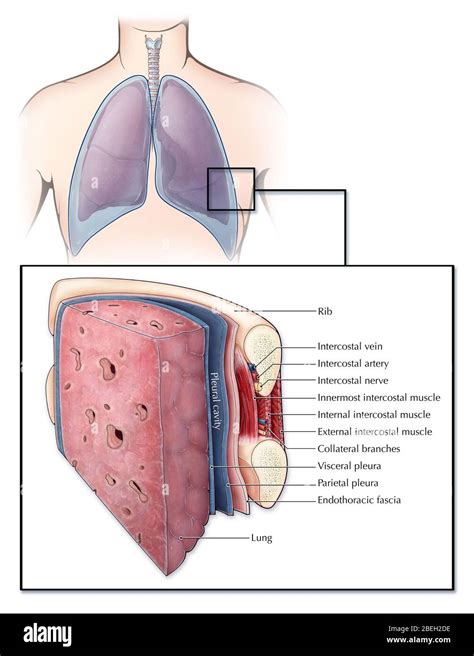
Table of Contents
The Visceral Pleura: The Inner Serosa Membrane Adhering to the Lungs
The lungs, the vital organs responsible for gas exchange, are not simply free-floating within the thoracic cavity. Instead, they are enveloped by a thin, double-layered serous membrane known as the pleura. This article delves deep into the intricacies of the visceral pleura, the inner layer of this membrane that intimately adheres to the lungs' surface. We'll explore its structure, function, and clinical significance, highlighting its crucial role in respiration and overall lung health.
Understanding the Pleura: A Protective Double Layer
Before focusing on the visceral pleura itself, it's important to understand the pleura as a whole. The pleura consists of two continuous layers:
- Visceral Pleura: This is the inner layer, also known as the pulmonary pleura. As mentioned, it's directly attached to the lung surface, dipping into the fissures between the lobes. Its surface is incredibly smooth, allowing for frictionless lung expansion and contraction during breathing.
- Parietal Pleura: This is the outer layer, lining the thoracic cavity's inner surface, including the diaphragm, the mediastinum (the space between the lungs containing the heart and other structures), and the inner surface of the rib cage.
Between these two layers lies the pleural cavity, a potential space containing a small amount of serous fluid. This fluid acts as a lubricant, minimizing friction between the visceral and parietal pleura during respiratory movements. This friction reduction is crucial for efficient and effortless breathing. The integrity of this fluid layer is essential for proper lung function.
Microscopic Structure of the Visceral Pleura: A Closer Look
The visceral pleura is not just a simple sheet of tissue; its microscopic structure is complex and contributes significantly to its function. It's composed of several layers:
-
Mesothelial Layer: This is the outermost layer, composed of simple squamous epithelium. These flattened cells are responsible for producing the pleural fluid and maintaining the integrity of the pleural cavity. The mesothelial cells have specialized functions including fluid regulation, immune responses and even some tissue repair. Their structure facilitates the smooth gliding motion of the lung within the thorax.
-
Submesothelial Connective Tissue: Beneath the mesothelium lies a layer of connective tissue. This layer is relatively thin but contains a network of collagen and elastic fibers, blood vessels, lymphatic vessels, and nerves. This connective tissue provides structural support and helps anchor the visceral pleura to the lung parenchyma (the functional tissue of the lung). The elastic fibers play a key role in accommodating the lung’s volume changes during respiration.
-
Adventitial Layer: In some areas, particularly where the visceral pleura adheres more tightly to the lung tissue, an adventitial layer can be found. This layer blends seamlessly with the lung's connective tissue, providing a strong attachment.
The Crucial Role of the Visceral Pleura in Respiration
The visceral pleura's smooth surface and its close adhesion to the lung are paramount to efficient respiration. The negative pressure within the pleural cavity, created by the opposing forces of the chest wall and the lung's elasticity, is vital for lung expansion. This negative pressure pulls the visceral pleura, and consequently, the lung tissue outwards, facilitating inhalation. During exhalation, the pressure equalizes, and the lung passively recoils to its resting state.
Any disruption to the visceral pleura's integrity can severely compromise this delicate balance, leading to impaired respiratory function. For example, inflammation or scarring can cause adhesions, restricting lung movement and impacting breathing capacity.
Clinical Significance of Visceral Pleura Conditions
Several clinical conditions directly involve the visceral pleura, significantly impacting lung function and overall health.
Pleuritis (Pleurisy): Inflammation of the Pleura
Pleuritis, also known as pleurisy, is an inflammation of the pleura, which can affect both the visceral and parietal layers. This inflammation results in pain, typically sharp and stabbing, worsened by deep breaths or coughing. The pain arises from the irritation of the nerve endings within the parietal pleura. Numerous causes can lead to pleuritis, including infections (viral, bacterial, fungal), autoimmune disorders, cancer, and pulmonary embolism.
Pleural Effusion: Fluid Buildup in the Pleural Space
Pleural effusion refers to an abnormal accumulation of fluid in the pleural cavity. This fluid can be transudative (low protein content, often due to heart failure or liver disease) or exudative (high protein content, indicating inflammation or infection). The excess fluid can compress the lung, hindering its expansion and reducing oxygen intake. Various conditions can cause pleural effusions, such as heart failure, pneumonia, cancer, and kidney disease. Thoracentesis, a procedure to remove fluid from the pleural cavity, is often necessary for diagnosis and treatment.
Pneumothorax: Collapsed Lung
A pneumothorax is a condition in which air enters the pleural cavity, causing the lung to collapse. This can occur spontaneously (spontaneous pneumothorax), be caused by trauma (traumatic pneumothorax), or be associated with underlying lung diseases. The presence of air disrupts the negative pressure within the pleural cavity, preventing the lung from fully expanding. Treatment often involves inserting a chest tube to remove the air and re-expand the lung. The visceral pleura plays a crucial role in the mechanism of pneumothorax as it is compromised and unable to maintain the negative intrapleural pressure.
Mesothelioma: A Rare and Aggressive Cancer
Mesothelioma is a rare and aggressive cancer that originates in the mesothelial cells of the pleura. It's strongly linked to asbestos exposure, with a long latency period between exposure and disease development. Mesothelioma is a devastating disease with a poor prognosis, and treatment options are limited. Early detection is crucial for maximizing treatment effectiveness.
Lung Cancer and the Visceral Pleura
Lung cancer can directly invade the visceral pleura, leading to pleural effusion and other complications. The spread of cancer cells to the pleura is a significant indicator of advanced disease. Pleural involvement can impact treatment decisions and prognosis.
Diagnostic Imaging and the Visceral Pleura
Several imaging techniques are essential for visualizing the visceral pleura and assessing its integrity. These include:
-
Chest X-ray: A chest X-ray is often the initial imaging modality used to evaluate pleural abnormalities. It can detect pleural effusions, pneumothorax, and other conditions affecting the pleura.
-
Computed Tomography (CT) Scan: CT scans provide more detailed images of the chest, allowing for better visualization of the pleural layers and any abnormalities. CT scans are particularly useful in assessing the extent of pleural involvement in lung cancer and other diseases.
-
Ultrasound: Ultrasound is a non-invasive technique that can be used to assess pleural effusions and guide thoracentesis.
Conclusion: The Unsung Hero of Respiration
The visceral pleura, while often overlooked, plays a critical role in maintaining healthy lung function. Its delicate structure and precise interactions with the parietal pleura and the lung parenchyma are essential for efficient respiration. Understanding the visceral pleura's anatomy, physiology, and clinical significance is vital for healthcare professionals involved in diagnosing and treating a range of respiratory conditions. Further research into the intricate functions of the visceral pleura, particularly the mesothelial cells, is likely to yield deeper insights into respiratory health and disease. Continued advances in diagnostic imaging and treatment strategies will hopefully improve outcomes for individuals affected by pleural-related conditions. The visceral pleura, though unseen and often unacknowledged, is a vital component of our respiratory system and deserves recognition for its silent but indispensable contribution to our well-being.
Latest Posts
Latest Posts
-
5 Weaknesses Of The Articles Of Confederation
Mar 19, 2025
-
In Texas Political Parties Help Candidates By Providing
Mar 19, 2025
-
A 59 Year Old Patient Is Reporting Difficulty Breathing
Mar 19, 2025
-
A Mathematical Phrase Containing At Least One Variable
Mar 19, 2025
-
What Is The Most Abundant Gas In The Atmosphere
Mar 19, 2025
Related Post
Thank you for visiting our website which covers about Inner Serosa Membrane That Adheres To The Lungs. . We hope the information provided has been useful to you. Feel free to contact us if you have any questions or need further assistance. See you next time and don't miss to bookmark.
