Label The Features Of A Neuromuscular Junction
Breaking News Today
Mar 15, 2025 · 5 min read
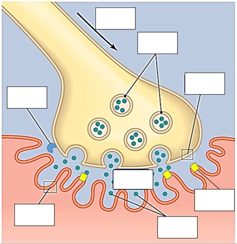
Table of Contents
Labeling the Features of a Neuromuscular Junction: A Comprehensive Guide
The neuromuscular junction (NMJ) is a specialized synapse where a motor neuron transmits a signal to a muscle fiber, initiating muscle contraction. Understanding its intricate structure and function is crucial for comprehending movement, neurological disorders, and various therapeutic interventions. This comprehensive guide will meticulously label and explain the key features of a neuromuscular junction, providing a detailed understanding of its complex machinery.
The Major Players: Motor Neuron and Muscle Fiber
Before delving into the specific components, it's vital to establish the two main actors:
-
Motor Neuron: A specialized nerve cell responsible for transmitting signals from the central nervous system (CNS) to skeletal muscle fibers. Its axon terminates at the NMJ, forming a synapse with the muscle fiber. The axon's terminal branches contain numerous synaptic vesicles filled with the neurotransmitter acetylcholine (ACh).
-
Muscle Fiber (Skeletal Muscle Cell): A long, cylindrical cell that constitutes skeletal muscle. Its specialized region, the motor endplate, receives the signal from the motor neuron and initiates the muscle contraction process.
Key Structural Features of the Neuromuscular Junction: A Detailed Breakdown
1. Axon Terminal (Presynaptic Terminal):
This is the very end of the motor neuron's axon. It's characterized by:
-
Synaptic Vesicles: Tiny membrane-bound sacs within the axon terminal, densely packed with acetylcholine (ACh). These vesicles are crucial for neurotransmission. Their release is triggered by the arrival of an action potential.
-
Voltage-Gated Calcium Channels: Located on the axon terminal membrane, these channels open when an action potential arrives, allowing calcium ions (Ca²⁺) to rush into the axon terminal. This influx of Ca²⁺ is the crucial trigger for exocytosis – the release of ACh from synaptic vesicles.
-
Mitochondria: Abundant within the axon terminal, these organelles provide the energy (ATP) required for the synthesis and release of ACh and for maintaining the overall function of the axon terminal.
-
Active Zones: Specialized regions within the presynaptic membrane where synaptic vesicles fuse and release their neurotransmitter. These zones ensure efficient and targeted neurotransmission.
2. Synaptic Cleft:
This is the narrow gap, approximately 20-30 nanometers wide, separating the axon terminal from the motor endplate of the muscle fiber. It's filled with extracellular matrix, and it's crucial for the diffusion of ACh across the synapse.
3. Motor Endplate (Postsynaptic Membrane):
This specialized region of the muscle fiber's membrane is located directly opposite the axon terminal. It's characterized by:
-
Junctional Folds: Deep invaginations of the motor endplate membrane significantly increasing the surface area available for ACh receptors. This amplification of the signal is vital for efficient muscle activation.
-
Nicotinic Acetylcholine Receptors (nAChRs): These ligand-gated ion channels are embedded in the motor endplate membrane. When ACh binds to these receptors, they undergo a conformational change, opening the channel and allowing the influx of sodium ions (Na⁺) into the muscle fiber. This depolarization initiates the muscle contraction process.
-
Acetylcholinesterase (AChE): This enzyme is located in the synaptic cleft and on the postsynaptic membrane. It rapidly hydrolyzes ACh, breaking it down into choline and acetate. This breakdown is essential for terminating the signal, preventing prolonged muscle contraction and allowing for precise control of muscle activity.
The Process of Neuromuscular Transmission: A Step-by-Step Guide
-
Action Potential Arrival: An action potential travels down the motor neuron axon to the axon terminal.
-
Calcium Influx: The arrival of the action potential triggers the opening of voltage-gated calcium channels, allowing Ca²⁺ to enter the axon terminal.
-
Synaptic Vesicle Fusion and ACh Release (Exocytosis): The increase in intracellular Ca²⁺ concentration triggers the fusion of synaptic vesicles with the presynaptic membrane, releasing ACh into the synaptic cleft.
-
ACh Binding to nAChRs: Released ACh diffuses across the synaptic cleft and binds to nAChRs on the motor endplate.
-
Sodium Influx and Depolarization: nAChR activation opens the ion channels, allowing Na⁺ to enter the muscle fiber. This influx of positive charge depolarizes the motor endplate, generating an end-plate potential (EPP).
-
Muscle Fiber Action Potential: The EPP triggers an action potential in the muscle fiber membrane. This action potential propagates along the muscle fiber, leading to muscle contraction.
-
ACh Breakdown: AChE rapidly hydrolyzes ACh in the synaptic cleft, terminating the signal and allowing the nAChRs to return to their resting state.
Clinical Relevance: Diseases Affecting the Neuromuscular Junction
Understanding the NMJ is critical for comprehending several neurological and muscular disorders. Dysfunction at any stage of neuromuscular transmission can lead to significant impairments. Here are a few examples:
-
Myasthenia Gravis: An autoimmune disease where antibodies attack and destroy nAChRs, leading to muscle weakness and fatigue.
-
Lambert-Eaton Myasthenic Syndrome (LEMS): Another autoimmune disease affecting voltage-gated calcium channels at the presynaptic terminal, impairing ACh release and causing muscle weakness.
-
Botulism: Caused by botulinum toxin, which blocks ACh release, resulting in paralysis.
-
Congenital Myasthenic Syndromes (CMS): A group of inherited disorders involving various components of the NMJ, leading to muscle weakness.
Advanced Concepts and Further Exploration
The NMJ is a dynamic and highly regulated structure. Further exploration might involve:
-
Synaptic plasticity: The ability of the NMJ to adapt and change its strength of transmission in response to activity.
-
Molecular mechanisms of ACh receptor clustering: The precise mechanisms that ensure the precise localization of nAChRs at the motor endplate.
-
The role of other neurotransmitters and neuromodulators: The influence of other molecules on NMJ function.
-
Developmental aspects of NMJ formation: The intricate process by which the NMJ is formed during development.
Conclusion
The neuromuscular junction is a fascinating and complex structure essential for voluntary movement. This detailed labeling and explanation of its features provide a solid foundation for understanding its function and its role in health and disease. By comprehending the intricacies of neuromuscular transmission, we can gain valuable insights into neurological disorders and develop more effective treatments. Further research continues to unveil the complexity and adaptability of this critical synapse, offering new avenues for therapeutic advancements.
Latest Posts
Latest Posts
-
What Should You Do With The Contact Trip Trigger
Mar 15, 2025
-
The Presidency Of Bill Clinton Quick Check
Mar 15, 2025
-
Why Are Coastal Areas A Focus Of Conservation Efforts
Mar 15, 2025
-
Driving A Vehicle On Public Roads Is Best Described As
Mar 15, 2025
-
In The Term Rhonchus The Root Rhonch Means
Mar 15, 2025
Related Post
Thank you for visiting our website which covers about Label The Features Of A Neuromuscular Junction . We hope the information provided has been useful to you. Feel free to contact us if you have any questions or need further assistance. See you next time and don't miss to bookmark.
