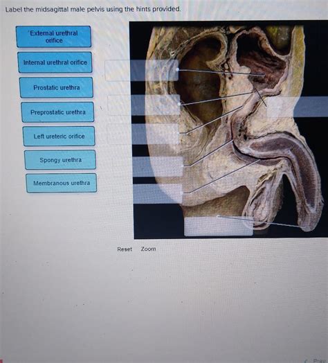
Label The Midsagittal Male Pelvis Using The Hints Provided
Breaking News Today
Apr 04, 2025 · 6 min read

Table of Contents
Label the Midsagittal Male Pelvis: A Comprehensive Guide
The male pelvis, a complex structure crucial for support and locomotion, presents a unique anatomical arrangement. Understanding its intricate components is vital for medical professionals, students, and anyone interested in human anatomy. This guide will walk you through labeling a midsagittal view of the male pelvis, utilizing hints and detailed descriptions to enhance your understanding. We'll cover key bony landmarks, ligaments, and muscles associated with this critical region.
Understanding the Midsagittal Plane
Before we dive into labeling the pelvis, let's establish a clear understanding of the midsagittal plane. This vertical plane divides the body into equal right and left halves. A midsagittal view of the pelvis provides a clear, side-on profile, showcasing the structures' depth and relationships. This perspective is particularly helpful for understanding pelvic dimensions and the articulation of bones.
Key Bony Landmarks of the Male Pelvis: A Step-by-Step Guide
The male pelvis differs subtly from the female pelvis, exhibiting characteristics reflecting its role in supporting greater weight and facilitating different biomechanical functions. Let’s systematically label the key bony landmarks:
1. Sacrum:
- Hint: This large, triangular bone forms the posterior wall of the pelvis. It's formed by the fusion of five sacral vertebrae.
- Labeling: Locate the sacrum; it's easily identifiable by its wedge-like shape and prominent anterior and posterior surfaces. Note the sacral foramina (openings for nerves and blood vessels) on its lateral aspects. Identify the sacral promontory, the anterior projection of the superior border of the S1 vertebra. The apex of the sacrum articulates with the coccyx.
2. Coccyx:
- Hint: This small, triangular bone at the very bottom of the spine is often described as the "tailbone."
- Labeling: Located inferior to the sacrum, the coccyx is a fused remnant of the four coccygeal vertebrae. Its articulation with the sacrum is typically quite mobile.
3. Ilium:
- Hint: The largest portion of the hip bone, it forms the superior part of the pelvis. It has a characteristic wing-like shape.
- Labeling: The ilium's superior border, known as the iliac crest, is easily palpable. Identify the anterior superior iliac spine (ASIS) and the posterior superior iliac spine (PSIS), important landmarks for palpation and anatomical reference. Note the iliac fossa, the concave inner surface of the ilium. The auricular surface, a roughened area on the posterior ilium, articulates with the sacrum forming the sacroiliac joint.
4. Ischium:
- Hint: This bone forms the inferior and posterior portions of the hip bone. It features a prominent ischial tuberosity.
- Labeling: Identify the ischial spine, a projection that is important in obstetrics. The ischial tuberosity, the large, roughened projection, is your sitting bone. The ischial ramus contributes to the inferior and anterior aspect of the hip bone.
5. Pubis:
- Hint: This bone forms the anterior portion of the hip bone. It articulates with the pubis of the opposite side.
- Labeling: Locate the superior and inferior pubic rami. The pubic symphysis, the cartilaginous joint connecting the two pubic bones, is located at the midline. Identify the pubic crest and the pubic tubercle.
6. Acetabulum:
- Hint: This is the deep, cup-shaped socket that articulates with the head of the femur (thigh bone).
- Labeling: The acetabulum is formed by the fusion of the ilium, ischium, and pubis. It is easily identified by its deep concavity and location on the lateral aspect of the hip bone.
7. Obturator Foramen:
- Hint: This is a large opening formed by the ischium and pubis.
- Labeling: The obturator foramen is a significant anatomical landmark, allowing passage for nerves and blood vessels.
Pelvic Ligaments: Crucial for Stability
The bony structure of the pelvis is reinforced by several crucial ligaments, providing stability to the sacroiliac joints and the pubic symphysis. Understanding these ligaments is crucial for comprehending pelvic biomechanics:
1. Sacrotuberous Ligament:
- Hint: This strong ligament runs from the sacrum and coccyx to the ischial tuberosity.
- Labeling: Identify its broad, triangular shape extending from the posterior sacrum to the ischial tuberosity. It provides significant support to the sacroiliac joint.
2. Sacrospinous Ligament:
- Hint: This ligament runs from the sacrum to the ischial spine.
- Labeling: Located medial to the sacrotuberous ligament, this ligament, although smaller, also contributes significantly to pelvic stability.
3. Anterior Sacroiliac Ligament:
- Hint: This ligament connects the anterior surfaces of the sacrum and ilium.
- Labeling: Find this ligament on the anterior aspect of the sacroiliac joint, contributing to joint stability, although less robust than the posterior ligaments.
4. Interosseous Sacroiliac Ligament:
- Hint: This strong ligament lies deep within the sacroiliac joint.
- Labeling: This ligament is not typically visible in a midsagittal view. Its significance is implied by the stability of the sacroiliac joint.
5. Pubic Symphysis Ligaments:
- Hint: These ligaments connect the two pubic bones at the pubic symphysis.
- Labeling: These ligaments are primarily composed of fibrocartilage, which provides a degree of flexibility and support for the pubic symphysis.
Muscles Associated with the Male Pelvis: Function and Location
Several muscles are closely associated with the male pelvis, contributing to locomotion, posture, and bowel/bladder control. While a midsagittal section doesn't show all muscle details, key muscle attachments to pelvic bones are visible:
1. Piriformis:
- Hint: This muscle originates on the anterior surface of the sacrum and inserts on the greater trochanter of the femur.
- Labeling: Though partially obscured in a midsagittal view, its origin on the sacrum may be partially visible. Its importance in hip rotation should be noted.
2. Coccygeus:
- Hint: This muscle forms part of the pelvic floor.
- Labeling: Its origin and insertion are best observed in a more detailed view.
3. Levator Ani:
- Hint: This group of muscles forms the major portion of the pelvic floor, supporting pelvic organs and aiding in defecation and urination.
- Labeling: Parts of the levator ani muscles may be visible. Understanding their role in pelvic floor support is key.
4. Obturator Internus:
- Hint: This muscle passes through the lesser sciatic foramen and contributes to hip external rotation.
- Labeling: Its path and origin/insertion are better understood in other anatomical views.
Clinical Significance: Why Labeling Matters
Accurate labeling of the midsagittal male pelvis is vital for several reasons:
- Medical Diagnosis: Understanding the anatomy is crucial for diagnosing pelvic injuries, fractures, and other pathologies. Radiological images often require precise anatomical knowledge for interpretation.
- Surgical Planning: Surgeons rely on a comprehensive understanding of pelvic anatomy to plan and execute successful procedures.
- Physical Therapy: Physical therapists use this knowledge to assess and treat pelvic floor disorders and other musculoskeletal conditions.
- Obstetrics and Gynecology: Understanding the female pelvic anatomy (similar but with key differences) is vital for managing pregnancy, childbirth, and related conditions.
Tips for Accurate Labeling
- Use high-quality anatomical diagrams: Choose clear, detailed diagrams that provide a clear depiction of the midsagittal section.
- Start with the bones: Begin by identifying the major bones and work your way to the smaller structures.
- Use color-coding: If possible, use different colors to label different structures.
- Practice: Repeated practice will significantly enhance your ability to identify and label the structures correctly.
By carefully following this guide and practicing regularly, you will develop a strong understanding of the midsagittal male pelvis and the numerous structures that contribute to its complexity and crucial role in the human body. Remember to consult multiple sources and anatomical references to enhance your knowledge. The information provided here is for educational purposes and should not be considered a substitute for professional medical advice.
Latest Posts
Latest Posts
-
To Safely Control The Traffic Flow And Protect
Apr 04, 2025
-
Which One Would Be Considered Critical Information
Apr 04, 2025
-
What Are Three Components Of Human Resource Planning
Apr 04, 2025
-
Which Two Properties Are Required For Every Field
Apr 04, 2025
-
Which Of The Following Makeup Procedural Steps Comes First
Apr 04, 2025
Related Post
Thank you for visiting our website which covers about Label The Midsagittal Male Pelvis Using The Hints Provided . We hope the information provided has been useful to you. Feel free to contact us if you have any questions or need further assistance. See you next time and don't miss to bookmark.
