Label The Reproductive Structures Of The Female Pelvis
Breaking News Today
Apr 06, 2025 · 6 min read
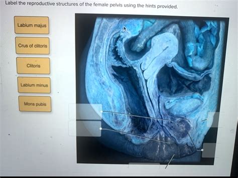
Table of Contents
Labeling the Reproductive Structures of the Female Pelvis: A Comprehensive Guide
Understanding the anatomy of the female reproductive system is crucial for healthcare professionals, students, and anyone interested in human biology. This detailed guide provides a comprehensive overview of the female pelvic reproductive structures, complete with clear labels and explanations. We'll explore each structure's function and its contribution to the overall reproductive process. This article aims to be a complete resource, covering all key aspects of female pelvic anatomy related to reproduction.
The Pelvic Cavity: The Foundation of Female Reproduction
The female pelvis, a bony structure, provides crucial protection and support for the reproductive organs. Its unique shape and size are essential for childbirth. Within this protective bony cage lies the intricate network of reproductive structures we will be exploring. Understanding the pelvic cavity's boundaries is the first step in understanding the location and relationships of these organs.
Bony Landmarks of the Pelvis:
- Iliac Crest: The superior border of the hip bone, easily palpable on the surface of the body.
- Pubic Symphysis: The cartilaginous joint connecting the two pubic bones at the front of the pelvis.
- Sacrum: A triangular bone formed by fused vertebrae at the back of the pelvis.
- Coccyx: The tailbone, a small, fused bone at the very end of the spinal column.
- Pelvic Inlet & Outlet: These define the boundaries of the pelvic cavity, crucial during childbirth.
These bony landmarks provide a framework for understanding the three-dimensional arrangement of the internal reproductive organs.
The Ovaries: The Source of Female Gametes
The ovaries are paired almond-shaped organs located on either side of the uterus, within the pelvic cavity. These are the primary female reproductive organs, responsible for:
- Oogenesis: The production of female gametes (ova or eggs). This process begins before birth and continues throughout a woman's reproductive years, albeit with a cyclical pattern.
- Hormone Production: The ovaries produce estrogen and progesterone, hormones critical for the development of secondary sexual characteristics, the menstrual cycle, and pregnancy. These hormones also influence various other bodily functions.
Microscopic Anatomy of the Ovary:
While we focus on the macroscopic structures here, it's important to acknowledge the microscopic complexity of the ovaries. Follicles, containing developing oocytes, are embedded within the ovarian stroma. The stages of follicle development, from primordial follicle to Graafian follicle, are intricate and essential to understanding oogenesis. The corpus luteum, a temporary endocrine gland, forms after ovulation and secretes progesterone.
The Fallopian Tubes (Uterine Tubes): The Pathway to Fertilization
The fallopian tubes, also known as uterine tubes or oviducts, are slender, paired tubes extending from the ovaries to the uterus. Their primary function is to transport the ovum from the ovary to the uterus.
- Infundibulum: The funnel-shaped opening of the fallopian tube near the ovary, featuring fimbriae (finger-like projections) that help capture the released ovum.
- Ampulla: The widest and longest part of the fallopian tube, where fertilization typically occurs.
- Isthmus: The narrower segment connecting the ampulla to the uterus.
- Interstitial Segment: The portion of the fallopian tube that passes through the uterine wall.
The cilia lining the fallopian tubes and peristaltic contractions propel the ovum towards the uterus. The journey takes approximately 3-4 days. If fertilization occurs within the ampulla, the fertilized ovum (zygote) begins its journey towards implantation in the uterus.
The Uterus: The Site of Implantation and Fetal Development
The uterus, a pear-shaped muscular organ, is located in the pelvic cavity between the bladder and the rectum. It's the primary site of fetal development during pregnancy.
- Fundus: The dome-shaped top portion of the uterus.
- Body (Corpus): The main part of the uterus.
- Isthmus: The constricted region between the body and the cervix.
- Cervix: The lower, narrow part of the uterus that extends into the vagina. The cervical canal connects the uterine cavity to the vagina.
Uterine Wall Layers:
The uterine wall consists of three layers:
- Perimetrium: The outer serous layer covering the uterus.
- Myometrium: The thick, muscular middle layer responsible for uterine contractions during labor and menstruation.
- Endometrium: The inner lining of the uterus, which undergoes cyclical changes during the menstrual cycle, preparing for potential implantation of a fertilized ovum. This layer is richly supplied with blood vessels.
The endometrium's cyclical changes are orchestrated by hormonal fluctuations, primarily estrogen and progesterone.
The Vagina: The Birth Canal and Copulatory Organ
The vagina is a fibromuscular canal extending from the cervix to the external genitalia. It serves as:
- Birth Canal: The passageway for childbirth.
- Copulatory Organ: The receptacle for the penis during sexual intercourse.
- Pathway for Menstrual Flow: The vagina allows for the expulsion of menstrual blood during menstruation.
The vaginal walls are lined with mucous membranes, which provide lubrication. The vaginal flora, a complex community of microorganisms, plays a crucial role in maintaining vaginal health. The acidic pH of the vagina helps to prevent infections.
Supporting Structures: Ligaments and Broad Ligament
Several ligaments provide support to the pelvic reproductive organs, keeping them in their proper anatomical position. The broad ligament, a large fold of peritoneum, encloses the fallopian tubes, ovaries, and uterus, providing support and housing blood vessels and nerves. Other ligaments, such as the ovarian ligament, suspensory ligament, and uterosacral ligament, play specific roles in supporting individual organs within the pelvic cavity.
The External Genitalia (Vulva): Protection and Sensory Input
The external genitalia, collectively known as the vulva, comprise:
- Mons Pubis: A fatty pad overlying the pubic symphysis, covered with pubic hair after puberty.
- Labia Majora: Two prominent folds of skin enclosing the other external genitalia.
- Labia Minora: Two smaller folds of skin located within the labia majora.
- Clitoris: A highly sensitive erectile organ, homologous to the penis.
- Vestibule: The area enclosed by the labia minora, containing the openings of the urethra and vagina.
- Bartholin's Glands: Located on either side of the vaginal opening, these glands secrete mucus for lubrication.
The external genitalia provide protection for the internal reproductive organs and contribute to sexual sensation.
Clinical Significance and Conclusion
Understanding the anatomy of the female reproductive system is paramount in various medical fields, including gynecology, obstetrics, and reproductive endocrinology. Accurate identification of these structures is essential for diagnosis and treatment of various conditions affecting the female reproductive tract, such as endometriosis, ovarian cysts, ectopic pregnancies, uterine fibroids, and sexually transmitted infections (STIs). Furthermore, knowledge of this intricate system allows for better understanding of reproductive health, family planning, and fertility treatments.
This comprehensive guide provides a solid foundation for understanding the reproductive structures of the female pelvis. Further study, including histological analysis and clinical experiences, will enhance understanding and allow for effective application of this knowledge in healthcare and related fields. Remember to consult reputable anatomical resources and textbooks for a more detailed and visually rich understanding of this fascinating system. This article serves as a starting point for a deeper dive into the intricacies of female reproductive anatomy and its importance in overall health.
Latest Posts
Latest Posts
-
Hair Design Is A Temporary Change In
Apr 08, 2025
-
Go Math Middle School Grade 7 Answer Key Second Edition
Apr 08, 2025
-
What Happens When Publicly Funded Youth Sport Programs Are Reduced
Apr 08, 2025
-
Experiment 23 Factors Affecting Reaction Rates Pre Lab Answers
Apr 08, 2025
-
Map Of Southeast Us States And Capitals
Apr 08, 2025
Related Post
Thank you for visiting our website which covers about Label The Reproductive Structures Of The Female Pelvis . We hope the information provided has been useful to you. Feel free to contact us if you have any questions or need further assistance. See you next time and don't miss to bookmark.