Label The Veins Of The Head And Neck
Breaking News Today
Mar 15, 2025 · 5 min read
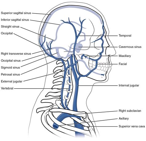
Table of Contents
Labeling the Veins of the Head and Neck: A Comprehensive Guide
Understanding the venous system of the head and neck is crucial for healthcare professionals, medical students, and anyone interested in human anatomy. This intricate network of vessels plays a vital role in returning deoxygenated blood from the brain, face, and neck to the heart. This comprehensive guide will delve into the detailed anatomy of these veins, providing a clear and structured approach to labeling them effectively. We'll cover the major veins, their tributaries, and key anatomical landmarks to aid in accurate identification.
Major Veins of the Head and Neck: A Systematic Approach
The venous drainage of the head and neck is complex, with multiple interconnected veins contributing to the overall system. We can broadly categorize these veins into those draining the superficial and deep structures. Understanding this distinction is fundamental to accurate labeling.
Superficial Veins: Draining the Skin and Subcutaneous Tissues
The superficial veins are primarily responsible for draining the skin and subcutaneous tissues of the head and neck. These veins are more readily visible and palpable compared to their deeper counterparts. Key superficial veins include:
-
Anterior Jugular Veins: These veins descend vertically along the anterior aspect of the neck, medial to the sternocleidomastoid muscles. They often anastomose (connect) with each other and with the external jugular veins. Remember: their location is crucial for proper identification.
-
External Jugular Veins: Located superficially along the lateral aspect of the neck, these veins are easily identifiable. They drain blood from the posterior scalp, auricle (ear), and face. Important Note: Variations in their course are common.
-
Posterior Auricular Veins: These smaller veins drain the posterior scalp and auricle. They typically join the external jugular vein.
-
Facial Veins: These veins run along the face, draining blood from the nose, cheeks, and lips. They have significant clinical importance due to their connections with intracranial venous sinuses. Key Feature: Look for their location alongside the facial artery.
-
Superficial Temporal Veins: These veins drain the scalp and temporal region of the head. They unite to form the retromandibular vein.
Deep Veins: Draining the Deeper Structures
The deep veins of the head and neck are responsible for draining the deeper structures, including the brain, muscles, and organs. These veins are less readily visible and require a deeper understanding of anatomical relationships. The key deep veins include:
-
Internal Jugular Vein: This is the largest vein of the neck, draining blood from the brain, face, and neck. It descends alongside the common carotid artery and vagus nerve within the carotid sheath. Crucial Point: It’s essential to note its relationship with these structures.
-
Vertebral Veins: These veins drain the vertebral column and its surrounding muscles. They eventually empty into the brachiocephalic veins. Consider: Their course follows the vertebral arteries.
-
Retromandibular Vein: This vein is formed by the union of the superficial temporal and maxillary veins. It further divides into anterior and posterior branches, eventually contributing to the internal jugular vein. Clinical Relevance: It plays a significant role in the drainage of the temporomandibular joint.
-
Pterygoid Plexus: A network of veins located deep within the infratemporal fossa, the pterygoid plexus drains blood from the masticatory muscles and other structures in this region. Important Consideration: Its complex anatomy makes it a challenge to label effectively.
Labeling Techniques and Best Practices
Effective labeling requires a systematic approach combining anatomical knowledge with visual aids. Here's a step-by-step guide:
-
Start with a High-Quality Anatomical Diagram: Begin with a clear and detailed anatomical diagram of the head and neck veins. Many online resources and textbooks provide excellent visuals.
-
Identify Key Landmarks: Before labeling any veins, identify key anatomical landmarks such as the sternocleidomastoid muscle, mandible, and mastoid process. These landmarks provide reference points for accurate vein identification.
-
Label the Major Veins First: Begin by labeling the largest and most prominent veins, such as the internal and external jugular veins. This provides a framework for labeling the smaller tributaries.
-
Label Tributaries and Connections: Once the major veins are labeled, focus on the smaller tributaries and their connections. Pay attention to the direction of blood flow.
-
Use Clear and Concise Labels: Use precise and unambiguous labels, avoiding any abbreviations that might lead to confusion. For instance, instead of "Ext. Jug.", write "External Jugular Vein."
-
Color-Coding (Optional): Using different colors for different vein groups can enhance clarity and visual organization. For example, superficial veins could be one color, while deep veins could be another.
Clinical Significance of Understanding Head and Neck Veins
A thorough understanding of the venous system of the head and neck is crucial for various clinical applications:
-
Venipuncture: Accurate knowledge of vein location is essential for successful and safe venipuncture procedures.
-
Surgical Procedures: Surgeons require precise anatomical knowledge to avoid injuring veins during head and neck surgeries.
-
Diagnosis and Treatment of Vascular Diseases: Understanding venous drainage patterns is crucial for diagnosing and managing conditions like deep vein thrombosis (DVT), varicose veins, and other vascular disorders.
-
Cancer Diagnosis and Treatment: Certain cancers may spread through the lymphatic system, and understanding the venous system is critical for cancer detection and treatment planning.
Advanced Concepts and Further Learning
For a more in-depth understanding, consider exploring these advanced topics:
-
Intracranial Venous Sinuses: This complex network of channels within the cranial cavity plays a crucial role in draining blood from the brain.
-
Venous Anastomoses: Understanding the connections and interrelationships between different veins is crucial for appreciating the redundancy and compensatory mechanisms within the venous system.
-
Embryological Development of Head and Neck Veins: Exploring the embryological development provides valuable insights into the complex arrangement of the adult venous system.
Conclusion: Mastering the Venous Anatomy of the Head and Neck
Mastering the labeling of head and neck veins requires consistent effort, meticulous attention to detail, and a thorough understanding of anatomical relationships. Utilizing high-quality anatomical diagrams, following a structured labeling approach, and seeking supplementary learning resources will enhance your knowledge and contribute to successful learning. This comprehensive understanding is essential for healthcare professionals, medical students, and anyone interested in delving deeper into the fascinating world of human anatomy. Remember that continuous learning and revisiting this complex anatomical area are key to retaining and mastering the information. Through dedicated study and practice, you can confidently label the veins of the head and neck, improving your understanding of this critical anatomical region.
Latest Posts
Latest Posts
-
Ap Bio Unit 8 Progress Check Mcq
Mar 19, 2025
-
Explain Why Scientists Believe That Warm Climates Provide Greater Biodiversity
Mar 19, 2025
-
The Following Is A Parking Regulation In California
Mar 19, 2025
-
Athletic Ability Will Always Guarantee Success In Sports
Mar 19, 2025
-
What Is The Authors Purpose For Including This Sentence
Mar 19, 2025
Related Post
Thank you for visiting our website which covers about Label The Veins Of The Head And Neck . We hope the information provided has been useful to you. Feel free to contact us if you have any questions or need further assistance. See you next time and don't miss to bookmark.
