Label The Veins Of The Upper Limb
Breaking News Today
Mar 15, 2025 · 7 min read
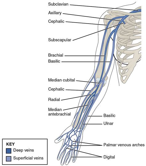
Table of Contents
Labeling the Veins of the Upper Limb: A Comprehensive Guide
Understanding the venous system of the upper limb is crucial for healthcare professionals, medical students, and anyone interested in human anatomy. This comprehensive guide will delve into the intricate network of veins in the arm, providing a detailed description, illustrations (imagine them here!), and practical tips for accurate labeling. We'll cover the superficial and deep veins, their tributaries, and their clinical significance. Mastering the labeling of these veins is key to understanding blood flow dynamics and diagnosing various vascular conditions.
Superficial Veins of the Upper Limb: A Detailed Look
The superficial veins are located just beneath the skin, making them readily accessible for venipuncture and other medical procedures. They're more variable in their anatomy than the deep veins, meaning their exact course and branching can differ between individuals. However, some key veins consistently appear. Let's explore these prominent superficial veins:
1. Cephalic Vein: The Lateral Route
The cephalic vein is the most lateral superficial vein of the upper limb. It originates on the radial side of the dorsum of the hand, running along the radial border of the forearm and then ascending along the lateral aspect of the arm. It eventually pierces the clavipectoral fascia and joins the axillary vein. It’s a significant vein often used for intravenous cannulation because of its superficial location and relatively consistent course. Remember: its lateral position is a key identifier.
2. Basilic Vein: The Medial Pathway
The basilica vein takes a more medial path. Originating on the ulnar side of the dorsum of the hand, it travels along the ulnar border of the forearm and then ascends along the medial aspect of the arm. Like the cephalic vein, it also eventually joins the axillary vein, usually after piercing the brachial fascia. Key difference: Its medial position distinguishes it from the cephalic vein.
3. Median Cubital Vein: The Connecting Link
The median cubital vein is a critical connecting vein, frequently bridging the cephalic and basilic veins in the cubital fossa (the anterior aspect of the elbow). This is the most common site for venipuncture due to its superficial location, large size, and relative stability. Important Note: Its position in the cubital fossa is a crucial landmark for both anatomical understanding and practical procedures.
4. Median Antebrachial Vein: A Variable Contributor
The median antebrachial vein is a variable vein located in the forearm. It often connects the cephalic and basilic veins, providing additional pathways for venous return. Its presence and exact course can vary significantly between individuals. Remember: its variability is a key characteristic.
5. Accessory Cephalic and Basilic Veins: The Supporting Cast
It's common to find accessory veins accompanying both the cephalic and basilic veins. These accessory veins provide additional drainage routes and contribute to the overall venous network complexity. Their presence and size vary, adding another layer of anatomical variability. Important Consideration: Always account for these variations when labeling.
Deep Veins of the Upper Limb: A Deeper Dive
The deep veins of the upper limb run alongside the major arteries, often sharing the same fascial compartments. They are usually paired veins, meaning two veins accompany each artery. These paired veins are crucial for efficient venous return. Let’s examine these deep venous structures:
1. Deep Palmar Arch and Dorsal Metacarpal Veins: The Hand's Foundation
The venous drainage of the hand begins with the deep palmar arch and the dorsal metacarpal veins. These veins collect blood from the digits and the hand, forming the initial stages of the deep venous system. They subsequently contribute to the more proximal veins of the forearm.
2. Radial and Ulnar Veins: Forearm's Primary Collectors
The radial vein and the ulnar vein are the primary deep veins of the forearm. They accompany the radial and ulnar arteries, respectively. These veins are responsible for collecting blood from the deep structures of the forearm, ultimately contributing to the brachial vein. Key Feature: Their paired nature mirrors the arterial system.
3. Brachial Vein: The Forearm's Culmination
The brachial vein is formed by the union of the radial and ulnar veins. This vein accompanies the brachial artery and is responsible for collecting blood from the entire forearm and carrying it towards the axilla. Remember: its formation signifies the transition from forearm to arm.
4. Axillary Vein: The Transition Point
As the brachial vein enters the axilla, it becomes the axillary vein. This vein receives tributaries from the shoulder and chest, playing a critical role in drainage from the upper limb. Crucial Location: Its position within the axilla indicates a change in anatomical region.
5. Subclavian Vein: The Final Leg of the Journey
Finally, at the lateral border of the first rib, the axillary vein becomes the subclavian vein. This marks the final segment of the upper limb's venous drainage, connecting the venous system of the arm to the larger venous network of the body. Key Transition: The subclavian vein signifies the end of the upper limb's venous system.
Clinical Significance and Practical Applications
Understanding the venous anatomy of the upper limb has crucial clinical applications:
-
Venipuncture: Accurate knowledge of superficial vein location is paramount for successful intravenous cannulation. The median cubital vein is frequently targeted due to its accessibility and size. However, understanding alternative veins is critical if the median cubital vein is unsuitable.
-
Thrombosis: Deep vein thrombosis (DVT) in the upper limb is less common than in the lower limb, but it can occur, particularly after trauma or surgery. Knowledge of the deep venous system is essential for accurate diagnosis and management.
-
Varicose Veins: Varicose veins, characterized by dilated and tortuous superficial veins, are more prevalent in the lower limbs but can affect the upper limb as well. Understanding superficial vein anatomy helps in diagnosis and treatment planning.
-
Lymphedema: Lymphedema, a condition characterized by swelling due to impaired lymphatic drainage, can affect the upper limb, often following breast cancer surgery or radiation therapy. Knowledge of the venous and lymphatic systems is essential for effective management.
-
Surgical Procedures: Surgical procedures involving the upper limb, such as vascular grafts or bypass surgeries, require a thorough understanding of the venous system for accurate surgical planning and execution.
Tips for Accurate Labeling: A Practical Guide
Accurate labeling requires a systematic approach. Here's a step-by-step guide to improve your labeling skills:
-
Start with a clear image or diagram: Use a high-quality anatomical image as your base. Ensure the image is large enough to allow for clear labeling.
-
Begin with the superficial veins: Label the cephalic, basilic, and median cubital veins first. Then, add the median antebrachial vein and any accessory veins.
-
Move to the deep veins: Label the radial, ulnar, and brachial veins. Ensure you correctly show their paired nature.
-
Transition to the axilla and clavicle: Label the axillary and subclavian veins, marking the transition points correctly.
-
Use concise and accurate labels: Avoid abbreviations unless they are widely understood. Ensure the labels are clearly positioned near the veins they identify.
-
Use different colors for superficial and deep veins (if possible): This can improve visual clarity and distinguish between the two systems.
-
Cross-reference your labels with anatomical texts: Use reliable anatomical texts to verify your labeling accuracy.
-
Practice regularly: Repeated labeling exercises are crucial for mastering the venous anatomy of the upper limb.
Conclusion: Mastering the Venous Network
Mastering the labeling of the veins of the upper limb requires diligent study and consistent practice. This guide provided a comprehensive overview of both the superficial and deep venous systems, highlighting their key features, clinical significance, and practical labeling techniques. Remember the variability inherent in human anatomy and always cross-reference your knowledge with reliable sources. With dedicated effort and a systematic approach, you will successfully navigate the complexities of this vital anatomical system. By understanding the intricacies of the upper limb veins, you lay the groundwork for a deeper understanding of human anatomy and its clinical implications.
Latest Posts
Latest Posts
-
Ap Bio Unit 8 Progress Check Mcq
Mar 19, 2025
-
Explain Why Scientists Believe That Warm Climates Provide Greater Biodiversity
Mar 19, 2025
-
The Following Is A Parking Regulation In California
Mar 19, 2025
-
Athletic Ability Will Always Guarantee Success In Sports
Mar 19, 2025
-
What Is The Authors Purpose For Including This Sentence
Mar 19, 2025
Related Post
Thank you for visiting our website which covers about Label The Veins Of The Upper Limb . We hope the information provided has been useful to you. Feel free to contact us if you have any questions or need further assistance. See you next time and don't miss to bookmark.
