Match Each Type Of Bone Marking With Its Definition
Breaking News Today
Mar 13, 2025 · 5 min read
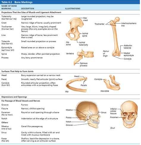
Table of Contents
Match Each Type of Bone Marking with Its Definition: A Comprehensive Guide
Bone markings, also known as surface features, are the various bumps, ridges, depressions, and openings found on the surface of bones. These aren't imperfections; they're crucial features that reflect the stresses placed upon the bone and reveal the points of attachment for muscles, ligaments, and tendons. Understanding these markings is essential for comprehending bone function, skeletal anatomy, and interpreting medical images like X-rays and CT scans. This comprehensive guide will meticulously define and explain each type of bone marking, providing examples where applicable.
Categories of Bone Markings
Before diving into specifics, it's helpful to categorize bone markings into broader groups:
1. Projections (Processes): These are areas that stick out from the bone surface. They serve as attachment points for muscles, tendons, and ligaments, or form joints with other bones.
2. Depressions (Cavities): These are indentations or openings in the bone. They often accommodate blood vessels, nerves, or other structures.
3. Openings: These are holes or channels that allow passage of blood vessels, nerves, or ligaments through the bone.
Detailed Breakdown of Bone Markings
Projections (Processes)
-
Condyle: A rounded, articular projection (a surface that forms a joint) that usually occurs in pairs. Think of the occipital condyles that articulate with the first vertebra (atlas).
-
Epicondyle: A projection situated above a condyle. The medial and lateral epicondyles of the humerus are prime examples, serving as attachment points for forearm muscles.
-
Facet: A small, smooth, flat articular surface. Vertebral facets are excellent illustrations, forming the joints between adjacent vertebrae.
-
Fissure: A narrow, slit-like opening. The superior orbital fissure in the skull allows passage of cranial nerves and blood vessels.
-
Foramen: A rounded passageway through a bone. The foramen magnum, the large opening at the base of the skull, is a crucial example, allowing the spinal cord to pass through.
-
Fossa: A shallow, basin-like depression. The mandibular fossa in the temporal bone is a significant example; it receives the mandible to form the temporomandibular joint.
-
Head: A prominent, rounded, articular end of a bone, often separated from the shaft by a narrower neck. The head of the femur is a classic example.
-
Crest: A prominent, narrow ridge of bone. The iliac crest, the superior border of the ilium, is a key landmark easily palpated on the hip.
-
Line: A slightly raised, elongated ridge of bone. The linea aspera on the femur provides attachment points for several thigh muscles.
-
Process: A projection or outgrowth of bone. This is a general term and encompasses many specific features, some of which we've already covered.
-
Protuberance: A bony outgrowth or projection. The chin is a prominent example, the mental protuberance.
-
Spine: A sharp, slender, pointed projection. The spine of the scapula is a very noticeable example.
-
Trochanter: A very large, blunt, irregularly shaped process. The greater and lesser trochanters of the femur are major sites of muscle attachment.
-
Tubercle: A small, rounded projection. The greater tubercle of the humerus is a crucial site for muscle attachment.
-
Tuberosity: A large, rounded projection. The tibial tuberosity is an excellent example, the site of patellar tendon attachment.
Depressions (Cavities)
-
Fovea: A small pit or depression. The fovea capitis is a small pit in the head of the femur where a ligament attaches.
-
Groove: A furrow or elongated depression. The intertubercular groove of the humerus accommodates the biceps brachii tendon.
-
Meatus: A tube-like passage or canal. The external acoustic meatus (ear canal) is an excellent example.
-
Sinus: A cavity within a bone. The paranasal sinuses are air-filled spaces within the skull bones, contributing to voice resonance and reducing skull weight.
-
Sulcus: A furrow, trench, or groove. Similar to a groove but often used to describe longer, more shallow depressions. The radial groove on the humerus accommodates the radial nerve and brachial artery.
Openings
-
Canal: A tunnel-like passageway. The haversian canals in compact bone allow passage of blood vessels and nerves.
-
Fissure: (already covered above as a projection but also functions as an opening)
Clinical Significance of Bone Markings
Understanding bone markings is crucial in various medical fields:
-
Orthopedics: Surgeons need to precisely identify bone markings during surgeries involving joint replacements, fracture repair, or tendon/ligament attachments. Misidentification can lead to complications.
-
Radiology: Radiologists interpret medical images like X-rays and CT scans, relying heavily on their knowledge of bone markings to identify fractures, dislocations, and other pathologies.
-
Anatomy and Physiology: A strong grasp of bone markings is paramount for students and professionals studying the musculoskeletal system.
-
Forensic Science: Bone markings can help forensic anthropologists determine the age, sex, and other characteristics of skeletal remains.
Advanced Considerations and Variations
The terminology surrounding bone markings can sometimes be ambiguous, with overlapping definitions and regional variations. Some sources may use different names for the same structure, or use the same name for slightly different structures. This is due to the complexity of the human skeleton and the historical evolution of anatomical terminology. It is crucial to rely on reputable anatomical texts and atlases for consistent and accurate information. Detailed anatomical study utilizing 3D models and anatomical specimens will help significantly in solidifying understanding.
Practical Applications and Further Exploration
To further solidify your understanding, try these activities:
-
Hands-on learning: Examine real bones (if accessible) or high-quality anatomical models, carefully identifying and labeling the various markings.
-
Image analysis: Study medical images (with appropriate supervision) to practice identifying bone markings in a clinical context.
-
Case studies: Research medical cases where misidentification of bone markings played a role in a diagnostic or surgical error (always from ethical and appropriately sourced materials).
-
Comparative anatomy: Explore the similarities and differences in bone markings between different species. This provides a richer understanding of functional adaptations.
This comprehensive overview has explored a wide array of bone markings, providing detailed definitions and examples. By mastering this information, you'll significantly enhance your understanding of human skeletal anatomy and its functional significance across various disciplines. Remember that continuous learning and practical application are crucial for deepening your knowledge and refining your skills in this area. The study of bone markings is an ongoing journey, constantly enriching our understanding of the human body's intricate design.
Latest Posts
Latest Posts
-
Which Part Of The Patient Record Is Classified As Administrative
May 09, 2025
-
A Recreational Flyer Is A Person Who
May 09, 2025
-
Tremors Hallucinations Delusions And Seizures Are All Symptoms Associated With
May 09, 2025
-
Which Internal Device Has The Largest Nonvolatile Storage Capacity
May 09, 2025
-
A Holder Of A Seller Server Certificate
May 09, 2025
Related Post
Thank you for visiting our website which covers about Match Each Type Of Bone Marking With Its Definition . We hope the information provided has been useful to you. Feel free to contact us if you have any questions or need further assistance. See you next time and don't miss to bookmark.