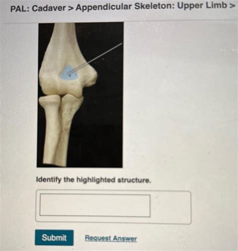
Pal Cadaver Appendicular Skeleton Upper Limb Lab Practical Question 3
Breaking News Today
Apr 04, 2025 · 6 min read

Table of Contents
Pal Cadaver Appendicular Skeleton Upper Limb Lab Practical Question 3: A Comprehensive Guide
This article delves deep into the complexities of the upper limb's appendicular skeleton, specifically addressing common practical questions encountered in anatomical labs using pal cadavers. We'll explore the bones, joints, and key anatomical features, providing detailed descriptions and practical tips for identification and understanding. This in-depth guide aims to enhance your understanding for lab practicals and beyond.
Understanding the Appendicular Skeleton of the Upper Limb
The appendicular skeleton of the upper limb consists of the bones of the arm, forearm, and hand. It's crucial to understand the individual bones, their articulations (joints), and their relative positions. Accurate identification requires meticulous observation and a systematic approach. Remember, the pal cadaver is a valuable tool, but careful handling and respect are paramount.
1. The Shoulder Girdle (Pectoral Girdle)
The shoulder girdle, comprised of the clavicle and scapula, forms the proximal attachment point for the upper limb to the axial skeleton.
-
Clavicle (Collarbone): A long, S-shaped bone. Observe its medial (sternal) end, articulating with the sternum, and its lateral (acromial) end, articulating with the acromion process of the scapula. Palpate for its characteristic curvature and note its palpable position just below the skin.
-
Scapula (Shoulder Blade): A flat, triangular bone located on the posterior thorax. Identify its prominent features:
- Acromion Process: The lateral extension forming the highest point of the shoulder. Its articulation with the clavicle forms the acromioclavicular joint.
- Coracoid Process: A curved process projecting anteriorly, providing attachment points for muscles.
- Glenoid Cavity: The shallow socket that articulates with the head of the humerus, forming the glenohumeral joint (shoulder joint). Note its relatively small size contributing to the shoulder's high mobility but decreased stability.
- Spine of the Scapula: A prominent ridge running across the posterior surface, terminating in the acromion process.
- Suprascapular Notch: A notch on the superior border, often incompletely formed, allowing passage for the suprascapular nerve.
2. The Free Upper Limb
The free upper limb consists of the humerus, radius, ulna, carpals, metacarpals, and phalanges.
-
Humerus: The long bone of the arm. Key features to identify include:
- Head: The rounded proximal end articulating with the glenoid cavity. Note its orientation and relationship to the anatomical neck.
- Greater and Lesser Tubercles: These bony projections provide attachment points for muscles. Locate their positions and note their relative size.
- Surgical Neck: A region distal to the tubercles, a common site of fracture.
- Deltoid Tuberosity: A roughened area on the lateral aspect, serving as the insertion site for the deltoid muscle. Palpate this region on yourself to understand its location.
- Capitulum and Trochlea: Distal articular surfaces articulating with the radius and ulna, respectively. Note the difference in their shapes.
- Medial and Lateral Epicondyles: Bony prominences on either side of the distal end, serving as attachments for forearm muscles.
-
Radius and Ulna: The two bones of the forearm.
- Radius: The lateral bone (thumb side). Observe its head articulating with the capitulum of the humerus and the radial notch of the ulna. Note the radial tuberosity, the site of biceps brachii muscle insertion. Its distal end is much larger than the proximal end.
- Ulna: The medial bone (pinky finger side). Identify its olecranon process (the point of your elbow), the coronoid process (anterior), and the trochlear notch articulating with the trochlea of the humerus. Its distal end is much smaller than the proximal end. Note the ulnar styloid process at the distal end.
3. The Hand
The hand's complex structure is comprised of:
-
Carpals (Wrist Bones): Eight small bones arranged in two rows. Accurate identification of individual carpals (scaphoid, lunate, triquetrum, pisiform, trapezium, trapezoid, capitate, hamate) requires careful study and practice. Focus initially on their relative positions and groupings. Note the palpable location of the scaphoid and pisiform.
-
Metacarpals (Palm Bones): Five long bones forming the palm. Identify their bases, shafts, and heads. Note the numbering system, with the first metacarpal being the thumb metacarpal.
-
Phalanges (Finger Bones): Each finger (except the thumb) has three phalanges: proximal, middle, and distal. The thumb has only two: proximal and distal. Observe their articulation and relative sizes.
Practical Tips for Lab Practical Success
Thorough preparation is key to excelling in your lab practical. Here's a structured approach:
-
Pre-Lab Study: Review relevant anatomical texts and diagrams. Familiarize yourself with the terminology and bone features. Create flashcards or diagrams to aid memorization.
-
Systematic Approach: Follow a consistent order when examining the pal cadaver. Begin with the larger bones and work your way to the smaller ones. Compare the left and right sides to identify any variations.
-
Palpation: Where possible, palpate the corresponding bones on your own body. This helps to correlate the cadaveric structures with your own anatomy.
-
Articulations (Joints): Pay close attention to the articulations between bones. Understand the types of joints (e.g., synovial, fibrous, cartilaginous) and their range of motion.
-
Muscle Attachments: While not the primary focus of this question, try to identify some major muscle attachment points on the bones. This will improve your holistic understanding of the upper limb's function.
-
Careful Handling: Remember that the pal cadaver represents a human being. Treat it with respect and handle it with care to prevent damage. Use appropriate tools and follow your instructor's guidelines.
-
Ask Questions: Don't hesitate to ask your instructor or teaching assistants if you have any questions or uncertainties. Clarifying doubts ensures a stronger understanding.
-
Practice Makes Perfect: Repetition is crucial. If possible, practice identifying the bones on multiple pal cadavers.
Common Mistakes to Avoid
Several common errors can hinder your lab practical performance:
-
Rushing: Take your time and carefully examine each bone. Rushing will lead to inaccuracies.
-
Lack of Preparation: Insufficient pre-lab study makes identification difficult.
-
Ignoring Details: Pay attention to subtle differences in bone shape and size. Small details can be crucial for accurate identification.
-
Poor Organization: A disorganized approach will make the task more difficult and increase the chances of error.
Beyond the Lab Practical: Clinical Relevance
Understanding the appendicular skeleton of the upper limb extends beyond the lab practical; it's crucial for various clinical applications. Knowledge of bone anatomy is essential for:
-
Diagnosis and Treatment of Fractures: Accurate identification of fracture sites requires a thorough understanding of bone morphology.
-
Surgical Procedures: Surgeons rely on detailed anatomical knowledge during operations involving the upper limb.
-
Orthopedic Assessments: Orthopedic specialists use their knowledge of bone anatomy to diagnose and treat musculoskeletal disorders.
-
Imaging Interpretation: Radiologists must be familiar with bone anatomy to interpret X-rays, CT scans, and MRIs.
Conclusion: Mastering the Upper Limb Appendicular Skeleton
This in-depth guide provides a comprehensive understanding of the pal cadaver appendicular skeleton, specifically focusing on the upper limb. By combining thorough pre-lab preparation, a systematic approach during the practical, and a focus on clinical relevance, you can confidently master the identification and understanding of the intricate structures within the upper limb. Remember, consistent effort and a commitment to learning will significantly contribute to your success in this vital area of anatomical study. The pal cadaver offers an unparalleled opportunity to build a strong foundation in human anatomy, setting you up for future success in your healthcare career.
Latest Posts
Latest Posts
-
In The Event Of A Skyjacking You Should Immediately
Apr 04, 2025
-
What Two Advantages Do Small Businesses Have Over Larger Companies
Apr 04, 2025
-
Which Of The Following Personally Owned Peripherals Cyber Awareness 2024
Apr 04, 2025
-
What Item Should Not Be Documented On A Performance Evaluation
Apr 04, 2025
-
The Eastern Bloc Became Dominated By The Soviets
Apr 04, 2025
Related Post
Thank you for visiting our website which covers about Pal Cadaver Appendicular Skeleton Upper Limb Lab Practical Question 3 . We hope the information provided has been useful to you. Feel free to contact us if you have any questions or need further assistance. See you next time and don't miss to bookmark.
