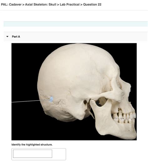
Pal Cadaver Axial Skeleton Skull Lab Practical Question 4
Breaking News Today
Apr 03, 2025 · 6 min read

Table of Contents
Pal Cadaver Axial Skeleton Skull Lab Practical: Question 4 and Beyond
This comprehensive guide delves into the intricacies of Question 4 in a typical pal cadaver axial skeleton skull lab practical, expanding beyond a simple answer to provide a thorough understanding of skull anatomy and its practical application. We will explore various aspects, including detailed bone identification, articulation, and clinical relevance, making this resource invaluable for students in anatomy, osteology, and related fields.
Understanding the Context: Pal Cadaver and Axial Skeleton
Before we tackle Question 4 specifically, let's establish the context. A "pal cadaver" refers to a preserved human body or body part used for anatomical study. In this instance, it's a skull, part of the axial skeleton. The axial skeleton is the central axis of the body, comprising the skull, vertebral column (spine), and rib cage. Understanding the relationship between these components is crucial for grasping the overall structure and function of the human skeleton.
The Importance of Hands-on Learning
Lab practicals using pal cadavers offer an unparalleled learning experience. Unlike textbook images or digital models, directly handling and examining real bone structures provides invaluable tactile and spatial understanding. This hands-on approach solidifies knowledge and improves identification skills, critical for future healthcare professionals.
Deconstructing Question 4: A Hypothetical Scenario
While the exact phrasing of Question 4 will vary depending on the specific lab manual, we can construct a hypothetical scenario to illustrate the typical challenges:
Hypothetical Question 4: "Identify the specific cranial bones contributing to the formation of the orbit (eye socket). Describe their articulations with one another and discuss the clinical significance of any fractures involving these bones."
Deep Dive into Cranial Bone Identification and Articulation
This question requires a detailed understanding of several key cranial bones and their intricate relationships:
1. Frontal Bone: The Forehead and Orbit's Roof
The frontal bone forms the anterior portion of the skull, including the forehead and the superior aspect of the orbit. Its contribution to the orbit forms the orbital roof. Identifying the frontal bone is relatively straightforward due to its smooth, curved surface and its prominent supraorbital ridges (brow ridges).
2. Zygomatic Bones: The Cheekbones and Orbital Walls
The zygomatic bones, or cheekbones, contribute significantly to the lateral and inferior walls of the orbit. Their strong, quadrilateral shape makes them readily identifiable. Note the zygomatic process, which articulates with the temporal bone to form the zygomatic arch.
3. Maxillae: The Upper Jaw and Orbital Floor
The maxillae form the upper jaw and contribute to the orbital floor (inferior wall). These bones are centrally located and articulate with numerous other bones. Carefully observe the infraorbital foramen, a significant landmark on the maxilla.
4. Sphenoid Bone: A Complex Bone with Orbital Involvement
The sphenoid bone is a complex, butterfly-shaped bone located deep within the skull. It contributes to the medial wall of the orbit via its lesser wings and body. Identifying the sphenoid requires a good understanding of its intricate structure.
5. Ethmoid Bone: Medial Orbital Wall and Nasal Cavity
The ethmoid bone, a delicate bone located between the orbits, contributes to the medial wall of the orbit. It also forms a significant portion of the nasal cavity. Its intricate structure includes the cribriform plate, olfactory foramina, and ethmoidal air cells.
6. Lacrimal Bones: Smallest Cranial Bones
The lacrimal bones are the smallest cranial bones. They are located in the medial wall of each orbit, posterior to the frontal process of the maxilla. Their small size and delicate nature require careful observation.
Articulations: Where Bones Meet
Understanding the articulations (joints) between these bones is crucial. These are primarily fibrous joints, characterized by strong connective tissue, providing stability and protection to the delicate orbital contents. The articulations are complex and often involve multiple bones.
Clinical Significance of Orbital Fractures
Orbital fractures can have severe consequences. The delicate structures within the orbit, including the eye, optic nerve, and blood vessels, are vulnerable to damage. The specific clinical presentation depends on the location and severity of the fracture.
Types of Orbital Fractures
-
Blow-out fractures: These are the most common type, involving the thin bones forming the orbital floor (maxilla) or medial wall (ethmoid). They occur when a blunt force is applied to the eye, causing the bone to fracture inward. This can trap the orbital contents, leading to diplopia (double vision), enophthalmos (sunken eye), and infraorbital nerve paresthesia.
-
Orbital rim fractures: These involve fractures of the bones surrounding the orbital rim (frontal, zygomatic, and maxilla). These fractures often present with visible facial deformities, periorbital ecchymosis (black eye), and possible nerve damage.
-
Zygomatic arch fractures: These fractures involve the zygomatic arch, compromising the lateral orbital wall. They are associated with significant facial deformity and malocclusion.
Beyond Question 4: Expanding Your Knowledge
Understanding Question 4 is a stepping stone to a deeper appreciation of the skull's complexity. Here are some additional areas to explore:
Sutures: The "Seams" of the Skull
The bones of the skull are joined together by fibrous joints called sutures. These sutures are named according to their location and the bones they connect. Understanding the different sutures helps in identifying individual bones and appreciating the overall cranial architecture. Examples include the coronal suture (between the frontal and parietal bones), the sagittal suture (between the parietal bones), and the lambdoid suture (between the parietal and occipital bones).
Foramina and Fissures: Passages for Nerves and Vessels
Numerous foramina (holes) and fissures (clefts) allow the passage of nerves, blood vessels, and other structures through the skull. Identifying these foramina and understanding their contents is critical in neuroanatomy and clinical practice. Examples include the optic canal (optic nerve), superior orbital fissure (oculomotor, trochlear, abducens, and ophthalmic nerves), and infraorbital foramen (infraorbital nerve).
Paranasal Sinuses: Air-Filled Cavities
The skull contains several air-filled cavities known as paranasal sinuses. These sinuses are located within the frontal, ethmoid, sphenoid, and maxillary bones. They contribute to the resonance of the voice and lighten the skull. Understanding their location and drainage pathways is essential in diagnosing sinus infections.
Clinical Correlations: Beyond Fractures
The knowledge gained from studying the skull's anatomy has far-reaching clinical applications beyond fracture management. Understanding cranial nerve pathways, blood supply, and the location of important structures is crucial in various medical specialties, including neurosurgery, ophthalmology, otolaryngology, and maxillofacial surgery.
Conclusion: Mastering the Pal Cadaver Skull Lab
The pal cadaver axial skeleton skull lab practical, including Question 4, provides an invaluable opportunity to solidify your understanding of cranial anatomy. By thoroughly studying the individual bones, their articulations, and clinical significance, you will develop a strong foundation for future studies in anatomy, physiology, and medicine. Remember that meticulous observation, accurate identification, and a comprehensive understanding of clinical relevance are crucial for success in this and any subsequent anatomical studies. This detailed exploration goes beyond a simple answer to Question 4, equipping you with a comprehensive understanding of the skull and its clinical implications. Remember to always approach the study of human anatomy with respect and a commitment to ethical practices.
Latest Posts
Latest Posts
-
Which Technology Was Originally Predicted By Science Fiction Writer
Apr 04, 2025
-
Gjaudro 1 Of 1 A Member Of A Team
Apr 04, 2025
-
Room Invasions Are Not A Significant Security Issue For Hotels
Apr 04, 2025
-
What Is True Of Malignant Melanoma Milady
Apr 04, 2025
-
Import Data From Classschedule Table In The Registration
Apr 04, 2025
Related Post
Thank you for visiting our website which covers about Pal Cadaver Axial Skeleton Skull Lab Practical Question 4 . We hope the information provided has been useful to you. Feel free to contact us if you have any questions or need further assistance. See you next time and don't miss to bookmark.
