Place The Images Of These Muscles In The Appropriate Category.
Breaking News Today
Apr 01, 2025 · 6 min read
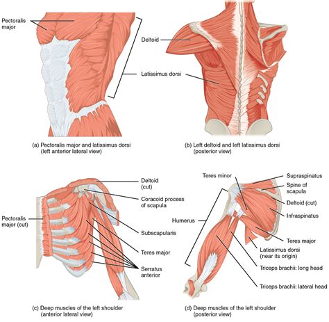
Table of Contents
Place the Images of These Muscles in the Appropriate Category: A Comprehensive Guide to Human Anatomy
Understanding human musculature is crucial for various fields, including medicine, physical therapy, fitness training, and art. This comprehensive guide will help you categorize images of muscles based on their location and function. We'll explore the major muscle groups of the body, providing detailed descriptions and visual cues to aid in accurate identification and categorization. This guide is designed to be both informative and practical, enhancing your understanding of human anatomy.
Categorizing Muscles: A Systematic Approach
Before we delve into specific muscle groups, let's establish a systematic approach to categorizing muscle images. We'll use a hierarchical system, dividing the body into major regions and then further subdividing these regions based on muscle function and location.
Primary Muscle Group Categories:
-
Head and Neck Muscles: These muscles control facial expressions, chewing, swallowing, and head movement. We'll further categorize these into facial muscles, masticatory muscles, and neck muscles.
-
Shoulder and Upper Extremity Muscles: This category encompasses muscles responsible for shoulder movement, arm flexion and extension, forearm rotation, and hand movements. Subcategories will include shoulder girdle muscles, arm muscles (anterior and posterior), forearm muscles (anterior and posterior), and hand muscles.
-
Thorax Muscles: Muscles of the chest and back, crucial for breathing and trunk stability. This will be broken down into muscles of respiration (intercostal muscles, diaphragm), and back muscles (superficial and deep).
-
Abdominal Muscles: Muscles forming the abdominal wall, essential for core stability and trunk flexion. These include the rectus abdominis, obliques, and transverse abdominis.
-
Pelvic Floor Muscles: These muscles support pelvic organs and are vital for continence and sexual function.
-
Lower Extremity Muscles: Muscles of the hip, thigh, leg, and foot, enabling locomotion and weight-bearing. We'll categorize them into hip muscles (anterior, posterior, medial), thigh muscles (anterior, posterior, medial), leg muscles (anterior, posterior), and foot muscles.
Detailed Muscle Group Analysis with Visual Cues
To effectively categorize muscle images, understanding the visual characteristics of each muscle group is paramount. We'll now examine each category in more detail, highlighting key features for identification:
1. Head and Neck Muscles
-
Facial Muscles: These are typically thin and superficial, responsible for a wide range of facial expressions. Look for their attachment points around the eyes (orbicularis oculi), mouth (orbicularis oris), and nose (nasalis). Images should show their delicate structure and often intricate fiber arrangements.
-
Masticatory Muscles: These muscles are involved in chewing. Key muscles to identify include the masseter (powerful, located on the side of the jaw), temporalis (fan-shaped, located above the ear), and pterygoids (deep, located within the jaw). Images will show strong, thick muscle bellies.
-
Neck Muscles: These muscles control head movement and posture. Look for the sternocleidomastoid (large, superficial, extending from the sternum and clavicle to the mastoid process), trapezius (broad, triangular, extending from the skull and spine to the clavicle and scapula), and other deeper neck muscles. Images may reveal their layered arrangement.
2. Shoulder and Upper Extremity Muscles
-
Shoulder Girdle Muscles: These muscles stabilize and move the scapula (shoulder blade). Key muscles to identify include the trapezius (already mentioned), levator scapulae, rhomboids (major and minor), and serratus anterior. Images may show their broad attachments and layered arrangement.
-
Arm Muscles (Anterior): Primarily responsible for elbow flexion. Look for the biceps brachii (two heads, easily recognizable), brachialis (deep to the biceps), and coracobrachialis. Images should show their distinct shapes and locations.
-
Arm Muscles (Posterior): Primarily responsible for elbow extension. The triceps brachii (three heads) is the dominant muscle here. Images will clearly show its three distinct heads.
-
Forearm Muscles (Anterior): These muscles are involved in wrist flexion, finger flexion, and pronation. Many are relatively small and deeply layered, making identification challenging without close examination of origin and insertion points.
-
Forearm Muscles (Posterior): These muscles are involved in wrist extension, finger extension, and supination. Again, many are small and layered. Careful observation of their attachment sites is crucial.
-
Hand Muscles: These are small and intricate muscles responsible for fine motor movements of the fingers and thumb. Images will need to be highly detailed to properly identify these muscles.
3. Thorax Muscles
-
Muscles of Respiration: The diaphragm is the primary muscle of breathing, forming a dome-shaped structure separating the thorax and abdomen. Intercostal muscles (external and internal) run between the ribs, assisting in breathing. Images should clearly show the diaphragm's structure and the intercostal muscles' direction of fibers.
-
Back Muscles (Superficial): Trapezius (already mentioned) and latissimus dorsi (broad, flat, extending from the spine to the humerus) are prominent superficial back muscles. Images will show their extensive attachments and layered positions relative to deeper muscles.
-
Back Muscles (Deep): These include erector spinae muscles (responsible for spinal extension), along with many smaller muscles involved in fine movements of the spine. Identifying these muscles requires detailed anatomical knowledge.
4. Abdominal Muscles
-
Rectus Abdominis: The "six-pack" muscle, extending vertically down the abdomen. Images should clearly show its segmented appearance.
-
External Obliques: These muscles run diagonally across the abdomen, forming a V-shape. Images will reveal their oblique fiber direction.
-
Internal Obliques: Located deep to the external obliques, their fibers run in the opposite direction.
-
Transverse Abdominis: The deepest abdominal muscle, running horizontally across the abdomen.
5. Pelvic Floor Muscles
These muscles are difficult to visualize directly in images without specialized anatomical preparations. However, understanding their general location and function is important. Key muscles include the levator ani (three parts: pubococcygeus, iliococcygeus, and puborectalis), coccygeus, and deep transverse perineal muscles.
6. Lower Extremity Muscles
-
Hip Muscles (Anterior): These muscles primarily flex the hip. Key muscles include the iliopsoas (iliacus and psoas major), sartorius, and rectus femoris (part of the quadriceps femoris).
-
Hip Muscles (Posterior): These muscles extend the hip. Gluteus maximus (large, superficial), gluteus medius, and gluteus minimus are key muscles here, along with the hamstrings (biceps femoris, semitendinosus, semimembranosus).
-
Hip Muscles (Medial): These muscles adduct the thigh. Adductor longus, adductor magnus, adductor brevis, and gracilis are important muscles in this group.
-
Thigh Muscles (Anterior): Quadriceps femoris (rectus femoris, vastus lateralis, vastus medialis, vastus intermedius) is the dominant muscle group, responsible for knee extension.
-
Thigh Muscles (Posterior): Hamstrings (already mentioned) are responsible for knee flexion and hip extension.
-
Thigh Muscles (Medial): These are mostly adductor muscles (already mentioned).
-
Leg Muscles (Anterior): Tibialis anterior, extensor hallucis longus, extensor digitorum longus are involved in dorsiflexion and toe extension.
-
Leg Muscles (Posterior): Gastrocnemius (superficial calf muscle), soleus (deep calf muscle), and tibialis posterior are involved in plantarflexion and inversion/eversion.
-
Foot Muscles: These small muscles are involved in fine movements of the toes.
Conclusion: Mastering Muscle Identification and Categorization
Successfully categorizing muscle images requires a combination of theoretical knowledge and practical observation. This guide has provided a structured framework, detailed descriptions of major muscle groups, and visual cues to help you accurately identify and categorize muscle images. By systematically reviewing this information and practicing with images, you can significantly enhance your understanding of human anatomy and build a strong foundation in musculoskeletal knowledge. Remember that practice makes perfect. The more images you analyze and categorize, the more proficient you will become in identifying and understanding the complex structure of the human muscular system. Consistent study and application of this knowledge will make you confident in your ability to accurately classify muscle images, regardless of the viewpoint or level of detail provided.
Latest Posts
Latest Posts
-
Liquid And Powder Nail Enhancements Are Created By
Apr 02, 2025
-
In Plants The Light Dependent Reactions Require
Apr 02, 2025
-
Three Legal Considerations In The Supervisory Relationship Are
Apr 02, 2025
-
Who Is Liable When An Insured Suffers A Loss
Apr 02, 2025
-
The San Andreas Fault In California Is An Example Of
Apr 02, 2025
Related Post
Thank you for visiting our website which covers about Place The Images Of These Muscles In The Appropriate Category. . We hope the information provided has been useful to you. Feel free to contact us if you have any questions or need further assistance. See you next time and don't miss to bookmark.
