The Authors Recommend An Exposure Sequence Starting With Tooth No.:
Breaking News Today
Mar 30, 2025 · 6 min read
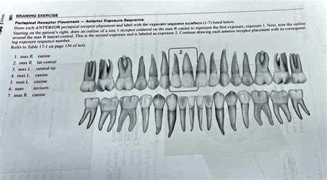
Table of Contents
The Authors Recommend an Exposure Sequence Starting with Tooth No.: A Comprehensive Guide to Dental X-Ray Techniques
Dental radiography is an indispensable tool in modern dentistry, providing invaluable insights into the health and condition of a patient's teeth and supporting structures. While the specific exposure sequence might vary slightly depending on the individual patient, the dental practice, and the available technology, a logical and efficient approach is crucial for both diagnostic accuracy and patient comfort. This article delves into the rationale behind recommended exposure sequences, focusing on the reasons for starting with a particular tooth number and the overall workflow involved. We will explore various factors influencing sequence selection and highlight best practices to ensure optimal image quality and minimize radiation exposure.
Why Start with a Specific Tooth? The Logic Behind Exposure Sequences
The question of "which tooth first?" isn't arbitrary. Several factors influence the preferred starting point for a full-mouth dental radiographic series. While there isn't a universally agreed-upon single "correct" starting tooth, the most common recommendations prioritize efficiency, anatomical landmarks, and radiation safety. Many authors and dental professionals recommend starting with either tooth #1 (the maxillary right first molar) or tooth #16 (the mandibular left first molar). Let's examine the reasons behind this:
1. Anatomical Landmarks and Workflow:
Starting with a maxillary molar (tooth #1 or #16) allows for a systematic approach, moving predictably through the arches. Maxillary molars frequently exhibit more complex anatomy, including multiple roots and furcations, requiring detailed radiographic assessment. Beginning here allows for a smooth transition through the maxilla and then the mandible, minimizing the need for significant repositioning of the X-ray unit and the patient. This streamlined workflow contributes to greater efficiency and reduced overall examination time.
2. Image Overlap and Positioning:
An effective sequence minimizes image overlap and ensures that all critical areas are captured clearly. Beginning with a molar and proceeding systematically often results in a more logical arrangement of films or digital sensors, preventing the need for repetitive repositioning and potentially improving image quality.
3. Radiation Safety:
While the cumulative radiation dose from a full-mouth series is relatively low, minimizing the patient's exposure time is always a priority. A well-planned exposure sequence enhances efficiency, which directly translates to a reduction in radiation exposure. A streamlined workflow avoids unnecessary repeats due to positioning errors or image overlap, further minimizing radiation.
4. Establishing a Baseline:
Starting with a molar often allows the dentist or radiographer to immediately establish a baseline assessment of the patient’s overall dental health. The condition of the molars can provide clues about potential periodontal disease, caries, or other issues, guiding the subsequent examination of the remaining teeth.
Different Exposure Sequences and Their Rationale
While starting with tooth #1 or #16 is a common recommendation, various sequences exist, each with its own rationale and advantages. These sequences usually fall into two main categories:
1. Posterior-Anterior Sequence:
This approach begins with the posterior teeth (molars and premolars), then progresses to the anterior teeth (incisors and canines). It's often preferred because it addresses areas prone to more complex pathology, like periodontal disease and extensive caries, earlier in the sequence. This allows for a comprehensive assessment of the posterior teeth before moving to the anterior region.
Advantages: Prioritizes posterior teeth, identifies potential problems early. Disadvantages: Might require more adjustments in X-ray unit positioning.
2. Anterior-Posterior Sequence:
This method starts with the anterior teeth and then moves to the posterior regions. It might be preferred when there's a focus on specific anterior issues like crowding, impacted canines, or orthodontic concerns.
Advantages: Focuses on anterior regions first if those are of primary concern. Disadvantages: May delay detection of significant posterior problems.
3. Quadrant-Based Sequences:
Some practitioners prefer a quadrant-based approach, where each quadrant of the mouth (maxillary right, maxillary left, mandibular right, mandibular left) is imaged systematically. This strategy offers excellent organization and is especially useful for patients with complex dental situations or significant restorations.
Advantages: Highly organized, easy to track individual quadrants. Disadvantages: May not be the most efficient method for routine examinations.
Factors Influencing the Choice of Exposure Sequence
The optimal exposure sequence isn't solely determined by a single factor. Instead, the dentist or radiographer considers several variables:
- Patient's dental condition: A patient with extensive periodontal disease might benefit from a sequence prioritizing the posterior teeth.
- Specific clinical questions: If a specific area is of concern, the sequence might be modified to prioritize that region. For example, a suspected impacted canine would necessitate a sequence emphasizing the anterior maxilla.
- Type of imaging system: Digital sensors often allow for a more flexible approach compared to conventional film-based methods.
- Patient cooperation: The patient's ability to comfortably remain still throughout the procedure influences the sequence's speed and efficiency.
- Time constraints: A busy practice might favor a sequence that is inherently fast and efficient.
Best Practices for Optimal Image Quality and Radiation Safety
Regardless of the chosen exposure sequence, several key principles ensure optimal radiographic images while minimizing radiation exposure:
- Proper Patient Positioning: Accurate positioning is paramount. Incorrect placement can lead to image distortion, blurring, and the need for retakes, increasing radiation exposure.
- Correct Beam Alignment: Ensuring the X-ray beam is properly aligned with the sensor or film is essential for sharp, diagnostic images. Incorrect alignment can result in elongation or foreshortening of the teeth.
- Appropriate Exposure Settings: Using the correct kVp (kilovoltage peak) and mA (milliamperage) settings is crucial. These settings must be adjusted according to the patient's anatomy and the type of radiograph being taken.
- Collimation: Using a collimator restricts the size of the X-ray beam, minimizing unnecessary radiation exposure to the patient.
- Digital Imaging Techniques: Digital radiography offers significant advantages in terms of radiation reduction, image manipulation, and storage compared to film-based methods.
- ALARA Principle: Dental professionals should always adhere to the ALARA principle ("As Low As Reasonably Achievable"), minimizing patient radiation exposure without compromising image quality.
Conclusion: A Tailored Approach to Dental Radiography
The optimal exposure sequence for dental radiography is not a "one-size-fits-all" solution. It’s a dynamic decision tailored to the individual patient’s needs, considering their dental history, specific clinical questions, and the available technology. While many authors and practitioners recommend beginning with tooth #1 or #16, the fundamental principles of efficiency, clear anatomical visualization, and minimizing radiation exposure should always guide the choice. Through a systematic and well-planned approach, dentists can obtain high-quality radiographs while ensuring the utmost safety for their patients. By mastering these techniques and understanding the underlying rationale, dental professionals can maximize the diagnostic potential of radiography and deliver optimal patient care. Continued education and adherence to best practices are crucial in this ever-evolving field.
Latest Posts
Latest Posts
-
Create A Space Large Enough To Give You To Maneuver
Apr 01, 2025
-
Many Jacks Use Hydraulic Power True False
Apr 01, 2025
-
Job Specifications Are Often Referred To As
Apr 01, 2025
-
Which Of The Following Is An Example Of Indexing
Apr 01, 2025
-
King Uses The Check And Promissory Note Metaphors To
Apr 01, 2025
Related Post
Thank you for visiting our website which covers about The Authors Recommend An Exposure Sequence Starting With Tooth No.: . We hope the information provided has been useful to you. Feel free to contact us if you have any questions or need further assistance. See you next time and don't miss to bookmark.
