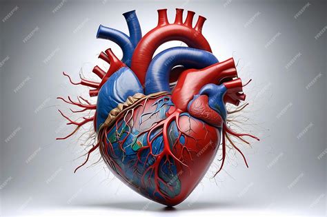
The Heart Is Medial To The Lungs
Breaking News Today
Mar 31, 2025 · 7 min read

Table of Contents
The Heart is Medial to the Lungs: An Anatomical Exploration
The statement, "the heart is medial to the lungs," is a fundamental concept in human anatomy. Understanding this spatial relationship is crucial for comprehending the intricate arrangement of organs within the thoracic cavity and for diagnosing and treating various medical conditions affecting the cardiovascular and respiratory systems. This article delves deep into this anatomical relationship, exploring the precise location of the heart and lungs, the implications of their positional arrangement, and the clinical significance of understanding this fundamental principle.
Defining Medial and Lateral in Anatomical Terminology
Before we delve into the specifics of the heart's position relative to the lungs, it's essential to clearly define the anatomical terms "medial" and "lateral." In anatomical terminology, medial refers to a structure being closer to the midline of the body, while lateral refers to a structure being further away from the midline. The midline is an imaginary vertical line that divides the body into equal left and right halves. Therefore, the heart being medial to the lungs signifies that it lies closer to the body's midline than the lungs do.
The Thoracic Cavity: A Protective Housing
The heart and lungs reside within the thoracic cavity, a protective space enclosed by the rib cage, sternum, and vertebral column. This cavity is further subdivided into compartments, allowing for the efficient and organized arrangement of vital organs. The lungs occupy the majority of the thoracic cavity's space, while the heart sits nestled within a more protected region known as the mediastinum.
The Mediastinum: A Central Compartment
The mediastinum is a central compartment within the thoracic cavity, separating the lungs into right and left lobes. It contains several crucial structures, including:
- The heart: The primary organ responsible for pumping blood throughout the body.
- The great vessels: Major blood vessels such as the aorta, vena cavae, and pulmonary arteries and veins.
- The trachea: The airway that carries air to and from the lungs.
- The esophagus: The tube that transports food from the mouth to the stomach.
- The thymus gland: An important component of the immune system.
- Nerves and lymph nodes: Contributing to the body's nervous and lymphatic systems.
The precise location of these structures within the mediastinum is meticulously regulated, ensuring optimal functioning and minimizing the risk of interference between different organ systems. The heart's position within the mediastinum, specifically its medial location relative to the lungs, is a key element of this organized arrangement.
The Heart's Position: More Than Just Medial
While stating that the heart is medial to the lungs is accurate, it provides a simplified picture. The heart's position is more complex and can be described using several anatomical terms:
- Medial: As previously explained, the heart is located closer to the midline than the lungs.
- Slightly left of midline: Although central in the mediastinum, the heart is tilted slightly towards the left side of the chest. This slight leftward tilt is significant, and it influences the location of heart sounds auscultated during physical examinations.
- Anterior: The heart is situated anteriorly (towards the front) within the thoracic cavity.
- Inferior: The apex (pointed end) of the heart points inferiorly (towards the bottom) and slightly to the left.
These additional positional descriptors provide a more comprehensive understanding of the heart's anatomical location within the chest, going beyond the simple medial relationship to the lungs. This precise positioning is crucial for the efficient functioning of the cardiovascular system.
Clinical Significance of Understanding the Heart's Position
Understanding the heart's medial position relative to the lungs and its overall placement within the thorax has significant clinical implications in several areas:
- Physical Examination: Accurate location of heart sounds during auscultation depends on knowing the heart's normal anatomical position. Deviations from the expected position might indicate underlying medical conditions such as displacement due to a large pleural effusion or mediastinal mass.
- Cardiovascular Imaging: Procedures like echocardiography, cardiac CT scans, and cardiac MRI rely on knowledge of the heart's position to correctly interpret images and diagnose conditions like heart valve disease, congenital heart defects, and myocardial infarction. The relationship between the heart and lungs is especially relevant in imaging modalities as the structures often appear adjacent to each other in scans.
- Surgical Procedures: Cardiac surgery necessitates precise knowledge of the heart's position and its relation to surrounding structures. The medial position relative to the lungs guides surgical approaches and minimizes the risk of iatrogenic injury to neighboring organs.
- Trauma Assessment: In cases of chest trauma, understanding the spatial relationship between the heart and lungs helps in assessing the extent of injury and prioritizing treatment. Penetrating injuries near the midline might affect the heart and its surrounding vessels more readily than injuries targeting the lungs laterally.
- Lung Conditions: Conditions like pneumothorax (collapsed lung) or pleural effusion (fluid accumulation in the pleural space) can displace the heart from its normal position. Recognizing this displacement is critical for diagnosis and treatment of the underlying lung condition.
This knowledge is paramount for precise surgical procedures, effective image interpretation, and the accurate diagnosis of both cardiac and pulmonary diseases.
The Interplay Between the Heart and Lungs
The heart and lungs have a close and interdependent relationship. Their proximity within the thoracic cavity is not just a matter of spatial arrangement; it reflects their functional integration in maintaining homeostasis.
Blood Supply and Oxygenation
The lungs are responsible for oxygenating the blood, while the heart circulates this oxygenated blood to the body’s tissues. This intricate interplay begins in the pulmonary circulation:
- Deoxygenated blood: Deoxygenated blood from the body returns to the right side of the heart.
- Pulmonary arteries: The heart pumps this deoxygenated blood to the lungs via the pulmonary arteries.
- Gas exchange: In the lungs, the deoxygenated blood releases carbon dioxide and picks up oxygen during gas exchange in the alveoli.
- Pulmonary veins: Oxygenated blood then returns to the left side of the heart via the pulmonary veins.
- Systemic circulation: The left side of the heart pumps this freshly oxygenated blood to the rest of the body via the aorta, initiating systemic circulation.
This continuous cycle of oxygenation and circulation highlights the fundamental interdependence of the heart and lungs, a relationship reflected in their close anatomical proximity within the thoracic cavity.
Neurological Control
The autonomic nervous system plays a vital role in regulating both cardiac and respiratory function. This system finely coordinates the actions of both organ systems to meet the body's changing metabolic demands. For example, during exercise, the autonomic nervous system increases both heart rate and respiratory rate to supply the increased demand for oxygen by working muscles. This coordinated response underscores the functional integration of the cardiopulmonary system.
Developmental Aspects
The development of the heart and lungs during embryogenesis is a complex process involving intricate interactions between various tissues and organs. The relative positioning of these organs is established early during development and maintains its essential spatial arrangement throughout life. Malformations during the development of these organs can result in severe congenital heart defects and lung anomalies, affecting the individual's cardiopulmonary function.
Conclusion
The statement that the heart is medial to the lungs is a seemingly simple anatomical fact. However, this statement forms the basis for a deeper understanding of the complex spatial relationships and functional integration of the cardiovascular and respiratory systems. This positional relationship is pivotal for various aspects of clinical practice, including physical examination, imaging interpretation, surgical procedures, and the assessment of cardiopulmonary disorders. Appreciating the precise location and relationship of the heart to the lungs and other structures in the mediastinum is crucial for healthcare professionals and anyone seeking a more comprehensive understanding of human anatomy and physiology. Understanding the medial location of the heart relative to the lungs is not only a foundational piece of anatomical knowledge but also a cornerstone for clinical practice and effective patient care.
Latest Posts
Latest Posts
-
Name The Area Pictured In The Hootsuite Mobile App
Apr 02, 2025
-
The Process Of Adapting Borrowed Cultural Traits
Apr 02, 2025
-
Act Vocabulary Crossword Puzzle 1 Answer Key
Apr 02, 2025
-
When Assessing An Elderly Male Who Complains Of Nausea
Apr 02, 2025
-
You Should Use Low Beams Whenever You Can
Apr 02, 2025
Related Post
Thank you for visiting our website which covers about The Heart Is Medial To The Lungs . We hope the information provided has been useful to you. Feel free to contact us if you have any questions or need further assistance. See you next time and don't miss to bookmark.
