The Primary Auditory Cortex Is Located In The
Breaking News Today
Mar 30, 2025 · 7 min read
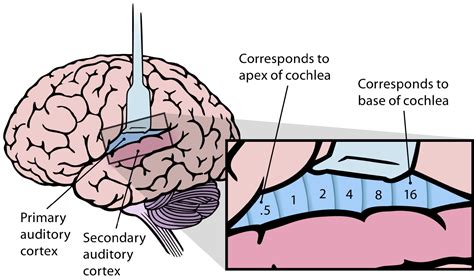
Table of Contents
The Primary Auditory Cortex: Location, Function, and Clinical Significance
The primary auditory cortex (A1) is the crucial initial processing center for auditory information in the brain. Understanding its precise location, intricate function, and clinical implications associated with damage or dysfunction is vital for comprehending the complexities of hearing and sound perception. This article delves deep into these aspects, providing a comprehensive overview for both students and professionals interested in neuroscience and audiology.
Precise Location of the Primary Auditory Cortex
The primary auditory cortex isn't a single, easily defined structure; instead, it's a collection of interconnected areas within the temporal lobe of the brain. More specifically, A1 resides within the superior temporal gyrus, a ridge of tissue located on the superior surface of the temporal lobe, just beneath the lateral sulcus (also known as the Sylvian fissure), a prominent groove that separates the temporal lobe from the frontal and parietal lobes.
Identifying A1: Cortical Mapping and Neuroimaging
Pinpointing the exact boundaries of A1 is challenging, even with advanced neuroimaging techniques. However, researchers have employed various methods to map its approximate location:
- Electrocorticography (ECoG): This invasive technique involves placing electrodes directly onto the surface of the brain, allowing for direct recording of electrical activity from specific cortical regions. ECoG studies have helped delineate the tonotopic organization of A1 (discussed in detail below).
- Functional Magnetic Resonance Imaging (fMRI): fMRI measures brain activity indirectly by detecting changes in blood flow. By presenting auditory stimuli and monitoring blood flow changes, researchers can identify brain areas involved in auditory processing, including A1.
- Magnetoencephalography (MEG): MEG measures magnetic fields produced by electrical activity in the brain. It offers a non-invasive way to identify the location and timing of neural activity in A1, providing high temporal resolution.
Despite these advancements, the precise borders of A1 remain somewhat debated. Its location is highly individual, varying slightly between individuals due to natural variations in brain anatomy. However, its consistent position within the superior temporal gyrus provides a reliable anatomical reference point.
Functional Organization: Tonotopy and Beyond
The primary auditory cortex isn't simply a passive receiver of auditory information; it's a highly organized structure with complex functional properties. One of the most striking features of A1 is its tonotopy.
Tonotopy: A Frequency Map in the Brain
Tonotopy refers to the systematic arrangement of neurons in A1 according to their characteristic frequencies. Neurons responding to low frequencies are located at one end of the cortex, while neurons responding to high frequencies are located at the other end. This orderly arrangement creates a “map” of sound frequencies across the cortex. This tonotopic organization isn't rigid; it's dynamic and can be modulated by experience and attention.
Beyond Tonotopy: More Complex Auditory Processing
While tonotopy is a cornerstone of A1's function, it’s only one piece of the puzzle. A1 also plays a role in processing other aspects of sound, including:
- Sound Intensity: Neurons in A1 exhibit varying sensitivity to sound intensity (loudness).
- Sound Location: While not the primary location for sound localization (that involves other brain areas), A1 contributes to the perception of sound location by integrating information from both ears.
- Temporal Aspects of Sound: A1 is sensitive to the timing of sounds, which is crucial for understanding speech and music.
- Spectral Complexity: A1 neurons respond differentially to the complex frequency patterns present in natural sounds.
The integration of these different aspects of sound information allows A1 to contribute significantly to the construction of our auditory percepts.
Connections and Interactions: A Network Perspective
The primary auditory cortex doesn't operate in isolation; it's deeply embedded within a larger network of auditory brain areas. Its intricate connections are crucial for its function:
Subcortical Connections: The Pathway to A1
Auditory information travels from the cochlea (inner ear) via the auditory nerve to the brainstem, then through several relay stations in the midbrain (cochlear nuclei, superior olivary complex, inferior colliculus), and finally to the medial geniculate body (MGN) in the thalamus. The MGN acts as a relay station, projecting information to the primary auditory cortex.
Cortical Connections: Beyond A1
A1 isn't the end of the auditory processing pathway. It's extensively interconnected with other cortical areas, including:
- Secondary Auditory Cortex (A2): A2, located adjacent to A1, processes more complex auditory features and contributes to sound recognition and categorization.
- Prefrontal Cortex: The prefrontal cortex is involved in higher-level cognitive processes related to auditory information, such as attention, memory, and decision-making.
- Other Sensory Areas: A1 interacts with other sensory areas, allowing for the integration of auditory information with visual and somatosensory information. This multimodal integration is crucial for our overall understanding of the world.
This intricate network of connections highlights the dynamic and interactive nature of auditory processing.
Clinical Significance: Consequences of A1 Damage
Damage to the primary auditory cortex, whether from stroke, trauma, or other neurological conditions, can have profound effects on auditory perception. The consequences aren't always straightforward, however, and depend on the extent and location of the damage within A1:
Cortical Deafness: A Rare but Severe Condition
In some cases, lesions encompassing a significant portion of A1 can result in cortical deafness. This is a rare condition characterized by difficulty processing auditory information, even in the absence of peripheral hearing loss. Individuals with cortical deafness may have normal hearing thresholds but struggle to discriminate sounds, understand speech, or localize sounds accurately.
Auditory Agnosias: Impaired Sound Recognition
Damage to A1 or interconnected areas can lead to auditory agnosias, which are characterized by difficulties recognizing familiar sounds despite intact hearing. Different types of auditory agnosia can exist, depending on the specific brain areas affected, for instance:
- Verbal Auditory Agnosia: Inability to understand spoken language.
- Non-Verbal Auditory Agnosia: Difficulty recognizing non-speech sounds like music or environmental noises.
Other Auditory Deficits: Beyond the Gross Impairments
Even relatively small lesions in A1 can result in more subtle auditory deficits. These might include:
- Difficulty discriminating similar sounds: For example, struggling to differentiate between similar phonemes in speech.
- Impaired sound localization: Difficulty pinpointing the source of a sound.
- Changes in sound perception: Alterations in the perceived loudness, pitch, or timbre of sounds.
The specific nature and severity of the deficits depend on a variety of factors including the precise location and extent of the lesion, the individual’s pre-existing auditory abilities, and the overall health of the brain.
Future Directions and Research
Research into the primary auditory cortex is an ongoing and dynamic field. Several exciting avenues of research are currently being explored:
Investigating Plasticity: The Brain's Adaptability
The brain exhibits remarkable plasticity, its ability to adapt and reorganize in response to experience or injury. Research is focused on understanding how A1 and its connected networks adapt following damage or alterations in auditory input.
Unraveling the Neural Code: How A1 Represents Sound
A fundamental question in auditory neuroscience concerns how the activity of neurons in A1 represents the acoustic features of sounds. Ongoing research utilizes advanced techniques such as multi-electrode recordings and computational modelling to unravel the neural code underlying sound perception.
Understanding Individual Differences: Personalized Audiology
The variability in auditory processing across individuals is increasingly being recognized. Research is focusing on identifying individual differences in A1 structure and function, which can inform the development of personalized approaches to hearing rehabilitation and treatment.
Conclusion
The primary auditory cortex is a critical component of the auditory system, playing a central role in processing sound information. Its precise location within the superior temporal gyrus, its intricate tonotopic organization, and its extensive connections with other brain areas are all crucial for our ability to hear and understand the world around us. Understanding the functional organization and clinical significance of A1 is essential for advancing our knowledge of auditory perception, developing effective treatments for hearing disorders, and creating personalized approaches to auditory rehabilitation. The ongoing research in this field promises to further our understanding of this fascinating and vital brain region.
Latest Posts
Latest Posts
-
First 36 Elements On The Periodic Table
Apr 01, 2025
-
A Computer Training Business Needs To Hire A Consultant
Apr 01, 2025
-
All Results Have An Obvious Link To A Landing Page
Apr 01, 2025
-
Identifying And Safeguarding Pii V4 Test Out Answers
Apr 01, 2025
-
Learning To Make Time An Ally Means
Apr 01, 2025
Related Post
Thank you for visiting our website which covers about The Primary Auditory Cortex Is Located In The . We hope the information provided has been useful to you. Feel free to contact us if you have any questions or need further assistance. See you next time and don't miss to bookmark.
