What Stage Of Cell Division Does This Image Show
Breaking News Today
Apr 01, 2025 · 6 min read
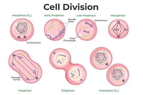
Table of Contents
What Stage of Cell Division Does This Image Show? A Comprehensive Guide
Determining the precise stage of cell division from an image requires careful observation and a solid understanding of the cell cycle. This article will provide a detailed walkthrough of identifying different stages, focusing on mitosis and meiosis, the two primary types of cell division. We'll explore the key characteristics of each stage, helping you confidently analyze microscopic images. While I cannot see the image you are referencing, I can guide you through the process of identification, focusing on the visual cues that differentiate each stage.
Understanding the Cell Cycle
Before diving into the stages, it's crucial to understand the broader context of the cell cycle. This cycle involves a series of precisely controlled events that lead to cell growth and division. The cycle is broadly divided into two major phases:
-
Interphase: This is the preparatory phase where the cell grows, replicates its DNA, and prepares for division. Interphase is further divided into G1 (gap 1), S (synthesis), and G2 (gap 2) phases. Microscopically, cells in interphase appear relatively uniform, with a clearly defined nucleus containing uncondensed chromatin.
-
M Phase (Mitotic Phase): This is the phase where cell division occurs. M phase encompasses mitosis (in somatic cells) or meiosis (in germ cells). Mitosis results in two genetically identical daughter cells, while meiosis produces four genetically diverse haploid gametes (sex cells).
Mitosis: A Detailed Look at the Stages
Mitosis, the process of somatic cell division, involves several distinct phases:
1. Prophase:
- Chromatin Condensation: The replicated DNA condenses into visible chromosomes. Each chromosome consists of two identical sister chromatids joined at the centromere. This is a crucial visual cue for identifying prophase. The nucleus is still largely intact at this stage, although the nuclear envelope may begin to break down.
- Spindle Fiber Formation: Microtubules begin to organize into a spindle apparatus, extending from the centrosomes (organelles that organize microtubules) toward the chromosomes.
- Nuclear Envelope Breakdown: Towards the late prophase, the nuclear envelope disassembles, allowing the chromosomes to interact with the spindle fibers.
2. Prometaphase:
- Chromosome Attachment: The spindle fibers attach to the kinetochores, protein structures located at the centromeres of each chromosome. This attachment is crucial for the accurate segregation of chromosomes during later stages.
- Continued Condensation: Chromosomes continue to condense and become more compact.
3. Metaphase:
- Chromosomal Alignment: The chromosomes align along the metaphase plate, an imaginary plane equidistant from the two poles of the spindle. This precise alignment is essential for ensuring that each daughter cell receives a complete set of chromosomes.
- Spindle Fiber Checkpoints: The cell undergoes a checkpoint to ensure that all chromosomes are correctly attached to the spindle fibers before proceeding to anaphase. This is a critical control mechanism preventing errors in chromosome segregation.
4. Anaphase:
- Sister Chromatid Separation: The sister chromatids separate at the centromere and are pulled towards opposite poles of the cell by the shortening spindle fibers. This is a dramatic visual change, with chromosomes appearing to be pulled apart.
- Chromosome Movement: The separated chromatids, now considered individual chromosomes, move toward opposite poles.
5. Telophase:
- Nuclear Envelope Reformation: The chromosomes arrive at the poles, and the nuclear envelopes begin to reform around each set of chromosomes.
- Chromatin Decondensation: The chromosomes begin to decondense, returning to their less compact chromatin form.
- Cytokinesis Initiation: Cytokinesis, the division of the cytoplasm, begins.
6. Cytokinesis:
- Cytoplasmic Division: The cell divides into two separate daughter cells, each containing a complete set of chromosomes. In animal cells, a cleavage furrow forms, pinching the cell in two. In plant cells, a cell plate forms, eventually developing into a new cell wall.
Meiosis: A Comparison with Mitosis
Meiosis is a specialized type of cell division that occurs in germ cells to produce gametes (sperm and egg cells). It involves two rounds of division: Meiosis I and Meiosis II. Each round has its own prophase, metaphase, anaphase, and telophase stages. The key differences lie in:
- Homologous Chromosome Pairing: In Meiosis I, homologous chromosomes (one from each parent) pair up during prophase I, forming bivalents. Crossing over, the exchange of genetic material between homologous chromosomes, occurs during this stage. This is a unique characteristic not seen in mitosis.
- Reductional Division: Meiosis I is a reductional division, reducing the chromosome number from diploid (2n) to haploid (n). This is achieved by separating homologous chromosomes, not sister chromatids, during anaphase I.
- Equational Division: Meiosis II is an equational division, similar to mitosis, where sister chromatids are separated.
Identifying the Stage from an Image: A Practical Approach
To determine the stage of cell division shown in an image, systematically analyze the following:
-
Chromosome Condensation: Are chromosomes visible? If not, the cell is likely in interphase. The degree of condensation helps distinguish between prophase (less condensed) and metaphase/anaphase (highly condensed).
-
Chromosome Arrangement: Are chromosomes aligned at the metaphase plate? This is a hallmark of metaphase. Are chromosomes moving toward opposite poles? This indicates anaphase.
-
Spindle Fibers: Are spindle fibers visible? Their presence and attachment to chromosomes are characteristic of stages from prometaphase onwards.
-
Nuclear Envelope: Is the nuclear envelope intact? Its absence suggests prometaphase or later stages.
-
Homologous Chromosome Pairing: If homologous chromosomes are paired, it strongly suggests meiosis I.
-
Sister Chromatid Separation: Are sister chromatids separating? This points towards anaphase (mitosis) or anaphase II (meiosis).
Examples of Microscopic Images and Their Interpretation
While I cannot see your specific image, let's consider hypothetical examples:
- Image showing uncondensed chromatin and a clearly defined nucleus: This strongly suggests interphase.
- Image showing condensed chromosomes but no alignment at the metaphase plate: This likely depicts prophase or prometaphase.
- Image showing chromosomes aligned at the metaphase plate: This is characteristic of metaphase.
- Image showing chromosomes moving toward opposite poles: This points towards anaphase.
- Image showing two distinct nuclei forming with decondensed chromosomes: This represents telophase followed by cytokinesis.
- Image showing paired homologous chromosomes: This is specific to Meiosis I, Prophase I.
Conclusion
Identifying the stage of cell division from a microscopic image requires a systematic approach and a thorough understanding of the cell cycle and its various stages. By carefully observing the characteristics of chromosomes, spindle fibers, and the nuclear envelope, you can accurately determine whether the cell is in interphase, mitosis, or meiosis, and pinpoint the specific stage within each process. Remember to consider the characteristics unique to meiosis, such as homologous chromosome pairing and the reductional division of Meiosis I. Practice analyzing various images, and you will quickly become proficient in interpreting these intricate cellular processes.
Latest Posts
Latest Posts
-
What Browser Must Be Used For Proctored Assignments
Apr 02, 2025
-
Harry Potter Order Of The Phoenix Ar Answers
Apr 02, 2025
-
Medication Fatigue Drugs Or Illness Can Tabc
Apr 02, 2025
-
This Is The Secret To Legendary Customer Service
Apr 02, 2025
-
What Principles Contribute To Personal And Professional Success
Apr 02, 2025
Related Post
Thank you for visiting our website which covers about What Stage Of Cell Division Does This Image Show . We hope the information provided has been useful to you. Feel free to contact us if you have any questions or need further assistance. See you next time and don't miss to bookmark.
