What Type Of Microbial Association Is Depicted In This Figure
Breaking News Today
Mar 22, 2025 · 6 min read
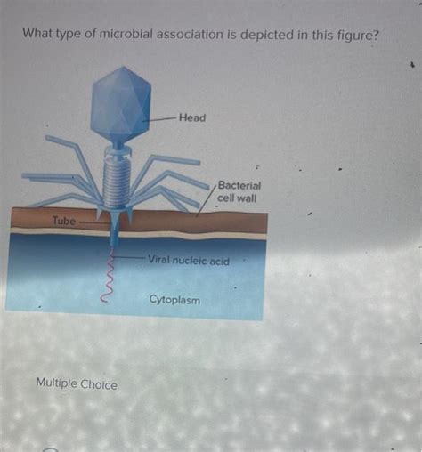
Table of Contents
Deciphering Microbial Associations: A Deep Dive into Figure Interpretation
This article delves into the interpretation of figures depicting microbial associations, offering a comprehensive guide for researchers and students alike. We'll explore various types of microbial interactions, focusing on visual identification within a figure context, and providing practical examples to solidify your understanding. The ultimate goal is to equip you with the skills to accurately identify and analyze the complex relationships within microbial communities based on visual representations.
Understanding Microbial Interactions: A Foundation
Before we analyze figures depicting microbial associations, let's establish a strong foundation by reviewing the fundamental types of interactions. Microbial communities are rarely composed of solitary organisms; instead, they engage in intricate relationships that shape their ecology and function. These relationships are broadly categorized as follows:
1. Mutualism: This is a win-win scenario where both interacting microbes benefit from the association. A classic example is the relationship between nitrogen-fixing bacteria (e.g., Rhizobium) and leguminous plants. The bacteria receive carbohydrates from the plant, while the plant gains access to essential nitrogen compounds fixed by the bacteria. In a figure, this might be represented by two organisms thriving in close proximity, with visual indicators suggesting resource exchange or mutual benefit.
2. Commensalism: In commensal relationships, one microbe benefits while the other is neither harmed nor helped. For instance, many bacteria living on human skin neither benefit nor harm the host, while potentially benefiting from a stable environment and access to nutrients. A figure depicting commensalism might show one microbe thriving near another without apparent interaction or influence.
3. Amensalism: Here, one microbe inhibits the growth or survival of another, without experiencing any reciprocal effect. This could be through the production of antibiotics or the depletion of essential resources. A figure might showcase one microbe thriving while the other shows signs of stress or decline in the presence of the first. The absence of direct interaction, but clear negative impact on one of the organisms, should be key indicators.
4. Parasitism: This is an antagonistic interaction where one microbe (the parasite) benefits at the expense of another (the host). Parasitism is often characterized by a dependency of the parasite on the host for survival and reproduction. Examples include bacteriophages infecting bacteria or pathogenic fungi infecting plant tissues. Visually, this might be depicted by one microbe visibly harming or damaging another, perhaps showing signs of structural damage or reduced growth.
5. Competition: This interaction occurs when two or more microbes compete for the same limited resources, such as nutrients or space. This often results in a negative impact on the growth or survival of at least one of the competitors. A figure might show two microbes in close proximity, with both exhibiting signs of reduced growth or abundance compared to when grown separately. Overlapping resource use zones could also be a visual cue.
6. Synergism: While not strictly a type of interaction like the others, synergism refers to a situation where two or more microbes working together achieve a combined effect that is greater than the sum of their individual effects. This often involves complementary metabolic activities or shared resource utilization. A figure depicting synergism might illustrate two microbes working collaboratively to perform a function that neither could achieve alone. The overall outcome – better growth or resource utilization – should be apparent.
Interpreting Figures: Visual Clues and Context
The key to accurately interpreting figures depicting microbial associations lies in observing several visual clues and carefully considering the provided context. Here's a breakdown of what to look for:
1. Spatial Proximity: Close proximity suggests a potential interaction, while distant organisms might have limited influence on each other.
2. Morphological Changes: Observe changes in the morphology (shape, size, structure) of the microbes involved. For instance, a deformed or damaged cell might indicate a parasitic or antagonistic interaction.
3. Abundance and Growth Rates: Compare the abundance and growth rates of microbes in different conditions or locations. A significant difference might hint at competition or amensalism.
4. Metabolic Byproducts: Figures might illustrate the presence or absence of metabolic byproducts indicating specific interactions. For example, the presence of certain enzymes or molecules associated with nitrogen fixation might indicate mutualistic symbiosis.
5. Co-localization: Do the microbes co-localize within a specific niche or habitat? Shared environments often indicate a degree of interaction.
6. Experimental Setup: The experimental setup is crucial. Figures representing co-cultures, where microbes are grown together, allow for the study of direct interactions. In contrast, figures depicting organisms grown in separate environments show individual characteristics without interference.
7. Staining and Microscopy Techniques: Different staining techniques might highlight specific structures or features relevant to interaction. Confocal microscopy and electron microscopy can provide high resolution images detailing cellular interactions.
8. Legend and Captions: The figure legend and captions are essential for proper interpretation. They provide context, explain the experimental methods, and identify the organisms involved.
Example Figure Interpretations: Putting it into Practice
Let's consider hypothetical scenarios and how we'd interpret them visually.
Scenario 1: A Figure Showing Two Bacterial Species Growing Together in a Co-Culture.
-
Observation: Both species show similar growth rates compared to individual cultures, exhibiting slight morphological changes. One bacteria species produces a particular enzyme, which allows the other to better utilize a certain nutrient.
-
Interpretation: This suggests a synergistic interaction; both species may improve their growth by working together. This collaborative metabolism enhances their overall efficiency and success.
Scenario 2: A Figure Depicting a Fungus Penetrating a Plant Root.
-
Observation: The fungal hyphae (thread-like structures) are penetrating the plant root cells, resulting in root damage and reduced plant growth.
-
Interpretation: This illustrates a parasitic relationship, where the fungus is deriving nutrients from the plant at its expense. The plant displays clear signs of harm and decreased fitness.
Scenario 3: A Figure Showing Two Bacterial Species Growing in Separate Zones Within the Same Habitat.
-
Observation: The two bacterial species occupy distinct zones and exhibit minimal overlap. Both species show healthy growth within their respective zones.
-
Interpretation: This might indicate a competitive interaction or niche partitioning. Both species are able to survive and thrive, but through segregation of resources they avoid direct confrontation.
Scenario 4: A Figure Showing Bacteria Growing on the Surface of a Mammalian Cell Without Apparent Harm.
-
Observation: The bacteria are attached to the surface of the mammalian cell, but there are no signs of cell damage or alteration. The bacteria appear to benefit from the stable environment provided by the host cell.
-
Interpretation: This may represent a commensal relationship; the bacteria benefit from the host, but the host is not demonstrably impacted.
Conclusion: The Importance of Critical Analysis
Interpreting figures depicting microbial associations requires a careful and critical approach. By understanding the different types of microbial interactions and looking for key visual clues within the provided context, researchers can accurately analyze the complex relationships within microbial communities. This understanding is essential for advancing our knowledge of microbial ecology, pathogenesis, and biotechnology. The ability to decipher these interactions from visual data is a critical skill for anyone working in the field of microbiology. Always remember to carefully examine the context, legend, and experimental setup to formulate a solid interpretation. The more practice you have interpreting such figures, the more confident and accurate your conclusions will be.
Latest Posts
Latest Posts
-
The Shape Of A Graduated Form Is
Mar 22, 2025
-
What Should You Not Do When Using A Stair Chair
Mar 22, 2025
-
The Stomach Is Inferior To The Diaphragm
Mar 22, 2025
-
What Type Of Rock Is Shown In This Photograph
Mar 22, 2025
-
A Basic Assumption Underlying The Definition Of Groupthink Is That
Mar 22, 2025
Related Post
Thank you for visiting our website which covers about What Type Of Microbial Association Is Depicted In This Figure . We hope the information provided has been useful to you. Feel free to contact us if you have any questions or need further assistance. See you next time and don't miss to bookmark.
