Which Valves Are Anchored By Chordae Tendineae
Breaking News Today
Apr 07, 2025 · 6 min read
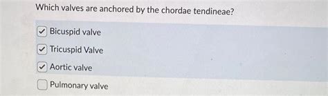
Table of Contents
Which Valves are Anchored by Chordae Tendineae? A Comprehensive Guide
The human heart, a marvel of engineering, relies on a complex interplay of chambers, valves, and supporting structures to efficiently pump blood throughout the body. One crucial component of this intricate system is the chordae tendineae, delicate but strong fibrous cords that play a vital role in the proper functioning of the heart valves. Understanding which valves these chordae tendineae anchor is fundamental to grasping the mechanics of the cardiovascular system and diagnosing potential heart conditions.
The Role of Chordae Tendineae in Heart Valve Function
Before delving into which valves are anchored, let's clarify the function of chordae tendineae. These tendon-like structures, also known as heart strings, are composed of collagen and elastin fibers. They originate from the papillary muscles, muscular projections within the ventricles (the heart's lower chambers). Their primary function is to prevent the atrioventricular (AV) valves from inverting or prolapsing during ventricular contraction (systole).
Imagine the AV valves as doors that need to open and close seamlessly to regulate blood flow. During ventricular contraction, the pressure inside the ventricles rises significantly. This pressure could force the AV valves open backward, allowing blood to flow back into the atria (the heart's upper chambers). The chordae tendineae, acting like guy wires, prevent this backflow by anchoring the valve leaflets (cusps) to the papillary muscles. This ensures unidirectional blood flow from the atria to the ventricles.
Identifying the Anchored Valves: Atrioventricular Valves
The answer to the question, "Which valves are anchored by chordae tendineae?" is straightforward: the atrioventricular (AV) valves. These valves are strategically positioned between the atria and ventricles, regulating the flow of blood between these chambers. There are two AV valves in the human heart:
1. The Mitral Valve (Bicuspid Valve)
Located between the left atrium and the left ventricle, the mitral valve is aptly named for its resemblance to a bishop's mitre. It possesses two leaflets (cusps) that open and close to regulate blood flow. These leaflets are firmly anchored to the papillary muscles of the left ventricle via the chordae tendineae. The robust connection ensures that the mitral valve doesn't prolapse during the forceful contraction of the left ventricle, maintaining the crucial one-way flow of oxygenated blood to the body.
Key features of mitral valve and chordae tendineae interaction:
- Robust attachment: The chordae tendineae connecting the mitral valve leaflets to the papillary muscles are numerous and strong, providing significant support.
- Precise coordination: The timing and strength of papillary muscle contraction are precisely coordinated with ventricular contraction to ensure effective valve closure and prevent regurgitation.
- Clinical significance: Mitral valve prolapse (MVP), a condition where the mitral valve leaflets bulge backward into the left atrium during ventricular contraction, often results from abnormalities in the chordae tendineae or papillary muscles.
2. The Tricuspid Valve
Positioned between the right atrium and the right ventricle, the tricuspid valve earns its name from its three leaflets (cusps). Similar to the mitral valve, these leaflets are also anchored by chordae tendineae that connect to the papillary muscles of the right ventricle. This arrangement prevents backflow of deoxygenated blood from the right ventricle into the right atrium during ventricular contraction.
Key features of tricuspid valve and chordae tendineae interaction:
- Three leaflets, multiple attachments: The three leaflets of the tricuspid valve each have numerous chordae tendineae attaching to multiple papillary muscles, creating a complex yet effective anchoring system.
- Lower pressure system: The right ventricle operates under lower pressure than the left ventricle, therefore the chordae tendineae and papillary muscles of the tricuspid valve may be slightly less robust than those of the mitral valve.
- Clinical relevance: Tricuspid regurgitation, a condition where blood flows back from the right ventricle to the right atrium, can be caused by damage to the tricuspid valve leaflets or their chordae tendineae attachments.
Valves Not Anchored by Chordae Tendineae: Semilunar Valves
It is crucial to understand that not all heart valves are anchored by chordae tendineae. The semilunar valves, namely the pulmonary valve and the aortic valve, do not have this support system. These valves are located at the outlets of the ventricles, separating the ventricles from the pulmonary artery (pulmonary valve) and the aorta (aortic valve).
1. The Pulmonary Valve
The pulmonary valve controls blood flow from the right ventricle to the pulmonary artery, which carries deoxygenated blood to the lungs for oxygenation. This valve has three semilunar cusps but lacks chordae tendineae. Its closure is primarily reliant on the pressure difference between the right ventricle and the pulmonary artery. During ventricular relaxation (diastole), the pressure in the right ventricle falls below the pressure in the pulmonary artery, causing the semilunar cusps to close passively.
2. The Aortic Valve
The aortic valve controls blood flow from the left ventricle to the aorta, the main artery supplying oxygenated blood to the body. Like the pulmonary valve, it consists of three semilunar cusps and is not anchored by chordae tendineae. Its closure also relies on the pressure difference between the left ventricle and the aorta. During diastole, the pressure in the left ventricle drops, allowing the aortic valve cusps to close passively, preventing backflow of blood into the left ventricle.
Clinical Significance of Chordae Tendineae and Valve Function
Understanding the crucial role of chordae tendineae in maintaining the integrity and proper function of the atrioventricular valves is paramount in the diagnosis and treatment of various cardiovascular conditions. Several clinical scenarios highlight their importance:
-
Mitral valve prolapse (MVP): This condition involves the stretching or bulging of the mitral valve leaflets into the left atrium during ventricular contraction. Often, it's associated with abnormalities in the chordae tendineae, such as elongation, rupture, or abnormal attachment. Symptoms can range from asymptomatic to significant heart palpitations, shortness of breath, and chest pain.
-
Tricuspid regurgitation: This condition involves the backflow of blood from the right ventricle into the right atrium during ventricular contraction. It can be caused by damage to the tricuspid valve leaflets, papillary muscles, or the chordae tendineae. Symptoms can range from mild to severe, depending on the severity of regurgitation.
-
Infective endocarditis: This severe infection of the heart valves can involve damage to the chordae tendineae, leading to valve dysfunction. Early diagnosis and treatment are crucial to prevent life-threatening complications.
-
Congenital heart defects: Congenital anomalies involving the chordae tendineae can cause various heart valve problems, requiring surgical intervention in many cases.
Conclusion: A Delicate Balance for Efficient Blood Flow
The chordae tendineae are essential components of the heart's intricate valve system. Their precise anchoring of the atrioventricular valves ensures unidirectional blood flow, preventing backflow and maintaining efficient cardiac output. Understanding their function and the consequences of their malfunction is vital for clinicians in the diagnosis and management of various heart conditions. The precise interaction between the chordae tendineae, papillary muscles, and AV valve leaflets underscores the remarkable complexity and efficiency of the human cardiovascular system. Further research continues to expand our knowledge of the biomechanics and clinical significance of this crucial anatomical structure. By appreciating the delicate balance maintained by these structures, we can better comprehend the importance of maintaining cardiovascular health.
Latest Posts
Latest Posts
-
Recycled Tires Are Frequently Turned Into
Apr 08, 2025
-
Which Of The Following Best Defines Dynamic Stretching
Apr 08, 2025
-
Arrange The Events In The Correct Chronological Order
Apr 08, 2025
-
A Life Insurance Policyowner Does Not Have The Right To
Apr 08, 2025
-
One Difference Between Arthur Mitchell And Michaela Depricne
Apr 08, 2025
Related Post
Thank you for visiting our website which covers about Which Valves Are Anchored By Chordae Tendineae . We hope the information provided has been useful to you. Feel free to contact us if you have any questions or need further assistance. See you next time and don't miss to bookmark.