X Ray Record Image Of The Spinal Cord
Breaking News Today
Apr 01, 2025 · 6 min read
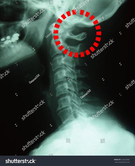
Table of Contents
X-Ray Record Images of the Spinal Cord: A Comprehensive Guide
X-ray imaging plays a crucial role in the diagnosis and management of spinal conditions. While not directly visualizing the spinal cord itself (as it's soft tissue and largely obscured by bone), X-rays provide invaluable indirect information about the bony structures that protect and support it. Understanding how to interpret these images is essential for healthcare professionals. This comprehensive guide explores various aspects of X-ray record images of the spinal cord, delving into the techniques, interpretations, and limitations.
Understanding the Limitations of X-Rays in Spinal Cord Imaging
Before delving into the specifics, it's crucial to acknowledge the inherent limitations of plain X-rays in visualizing the spinal cord. X-rays primarily reveal bone density differences. Soft tissues, including the spinal cord, nerves, and intervertebral discs, are largely transparent to X-rays and, therefore, not directly visible. Consequently, X-rays are not the primary imaging modality for evaluating the spinal cord itself. Conditions affecting the spinal cord are often better visualized using MRI, CT scans, or myelography.
However, X-rays are invaluable for assessing the bony structures that directly impact the spinal cord's health. These include:
- Vertebrae: X-rays clearly depict the shape, alignment, and integrity of the vertebrae. Fractures, dislocations, and degenerative changes are readily apparent.
- Intervertebral Foramina: These openings between vertebrae allow spinal nerves to exit the spinal canal. X-rays can reveal narrowing (stenosis) of these foramina, which can compress nerves and cause pain or neurological deficits.
- Spinal Canal: Although the spinal cord itself isn't visible, the size and shape of the spinal canal can be indirectly assessed. Narrowing or deformities of the canal can indicate potential compression of the spinal cord.
- Spinal Alignment: X-rays provide a clear picture of the overall alignment of the spine, revealing scoliosis (curvature), kyphosis (excessive forward curvature), and lordosis (excessive inward curvature).
Types of X-Ray Views for Spinal Imaging
Several standard X-ray views are employed to provide a comprehensive assessment of the spine. These views typically include:
- Anterior-Posterior (AP) View: This view is taken from the front to the back, providing a clear image of the vertebral bodies and their alignment. It's excellent for assessing vertebral fractures, dislocations, and the overall alignment of the spine.
- Lateral View: Taken from the side, this view is essential for evaluating the spinal curvature and the intervertebral spaces. It helps to visualize the size of the spinal canal and the presence of any abnormalities.
- Oblique Views: These views are taken at angles to the AP and lateral projections, providing supplementary information about the intervertebral foramina and the zygapophyseal joints (facet joints). They are particularly useful in assessing foraminal stenosis.
Interpreting X-Ray Images of the Spine: Key Findings
Interpreting spinal X-rays requires expertise and a systematic approach. Radiologists and other healthcare professionals trained in image interpretation look for several key findings:
Signs of Vertebral Fractures:
- Discontinuity of the cortical bone: A break in the outer layer of the bone is a clear indicator of a fracture.
- Loss of vertebral height: Compression fractures, common in osteoporosis, often result in a decrease in the height of the vertebral body.
- Displacement of vertebral fragments: In severe fractures, fragments of bone may be displaced from their normal position.
- Bone sclerosis: Increased bone density, often seen around the fracture site, indicates healing.
Signs of Degenerative Changes:
- Osteophytes: These bone spurs are common in degenerative joint disease and can narrow the intervertebral foramina.
- Disc space narrowing: A decrease in the height of the intervertebral disc space is indicative of disc degeneration.
- Facet joint osteoarthritis: Degeneration of the facet joints can lead to pain and stiffness.
- Subchondral sclerosis: increased bone density under the cartilage of the facet joints.
Signs of Spinal Alignment Abnormalities:
- Scoliosis: Lateral curvature of the spine.
- Kyphosis: Excessive forward curvature of the spine (e.g., dowager's hump).
- Lordosis: Excessive inward curvature of the spine (swayback).
- Spondylolisthesis: Forward slippage of one vertebra over another.
Signs of Spinal Stenosis:
- Narrowing of the intervertebral foramina: This can compress spinal nerves, causing pain and neurological symptoms.
- Narrowing of the spinal canal: This can compress the spinal cord, leading to myelopathy (spinal cord compression).
Advanced Imaging Techniques in Relation to X-Rays
While X-rays provide a foundational view, several advanced imaging techniques are often used in conjunction with them to obtain a more detailed understanding of spinal conditions. These include:
- Computed Tomography (CT) Scans: CT scans provide high-resolution images of bone and can reveal subtle fractures, dislocations, and other bony abnormalities that might be missed on plain X-rays. They are particularly useful in assessing complex fractures and spinal stenosis.
- Magnetic Resonance Imaging (MRI): MRI provides exceptional visualization of soft tissues, including the spinal cord, intervertebral discs, and spinal nerves. It's the preferred imaging modality for evaluating conditions affecting the spinal cord itself, such as spinal cord tumors, multiple sclerosis, and inflammatory diseases.
- Myelography: This involves injecting contrast material into the spinal canal, allowing for visualization of the spinal cord and its surrounding structures. It's often used in conjunction with CT (CT myelography) to better delineate spinal cord compression.
The Role of X-Ray Records in Patient Management
X-ray records of the spine serve as essential components of a patient's medical history. They are vital for:
- Diagnosis: X-rays help to diagnose a wide range of spinal conditions, from fractures and dislocations to degenerative diseases and spinal stenosis.
- Treatment Planning: The information provided by X-rays guides treatment decisions, determining the need for surgery, medication, or physical therapy.
- Monitoring Progress: Serial X-rays can be used to monitor the healing process after fractures or to assess the effectiveness of treatment for degenerative conditions.
- Legal Documentation: X-ray records provide objective documentation of a patient's spinal condition, which is often crucial in legal cases.
Conclusion
X-ray images of the spinal cord, while not directly visualizing the cord itself, offer invaluable information about the bony structures that protect and support it. They play a crucial role in diagnosing and managing a wide range of spinal conditions. Understanding the limitations of X-rays and their role in conjunction with advanced imaging techniques such as CT and MRI is vital for healthcare professionals involved in the diagnosis and treatment of spinal disorders. The interpretation of these images requires expertise and a systematic approach, focusing on key findings like fractures, degenerative changes, and spinal alignment abnormalities. Maintaining detailed and accurate X-ray records is crucial for effective patient management and long-term care. This comprehensive analysis highlights the significance of X-rays in providing a foundational understanding of spinal health. Further research and advancements in imaging technology will undoubtedly continue to refine our ability to diagnose and treat spinal disorders.
Latest Posts
Latest Posts
-
The Internet Is A Collection Of
Apr 02, 2025
-
When Testing Insulin Levels On Swimming Fish Hyperglycemia Results In
Apr 02, 2025
-
What Was One Immediate Result Of Henry Fords Manufacturing Methods
Apr 02, 2025
-
What Browser Must Be Used For Proctored Assignments
Apr 02, 2025
-
Harry Potter Order Of The Phoenix Ar Answers
Apr 02, 2025
Related Post
Thank you for visiting our website which covers about X Ray Record Image Of The Spinal Cord . We hope the information provided has been useful to you. Feel free to contact us if you have any questions or need further assistance. See you next time and don't miss to bookmark.
