A Sarcomere Is The Distance Between Two
Breaking News Today
Mar 17, 2025 · 7 min read
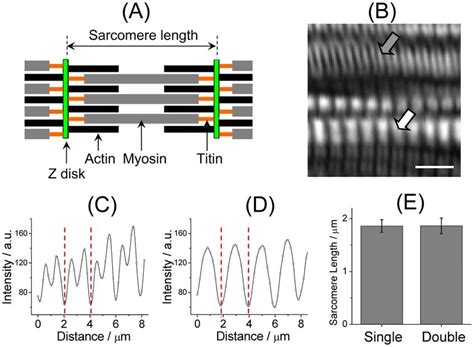
Table of Contents
A Sarcomere: The Distance Between Two Z-Lines – A Deep Dive into Muscle Contraction
The human body is a marvel of engineering, capable of feats of strength, endurance, and precision. At the heart of this capability lies the muscle, and within the muscle, the fundamental unit of contraction: the sarcomere. Understanding the sarcomere – defined as the distance between two Z-lines – is crucial to comprehending how muscles generate force and movement. This article will explore the sarcomere's structure, function, and the intricate mechanisms that govern muscle contraction.
The Sarcomere: A Microscopic Powerhouse
The sarcomere is the basic contractile unit of striated muscle tissue, such as skeletal and cardiac muscle. Its highly organized structure is responsible for the striated appearance seen under a microscope. This striation results from the precise arrangement of protein filaments within the sarcomere. Imagine the sarcomere as a highly sophisticated miniature machine, perfectly designed for generating force.
The defining feature of a sarcomere is the distance between two Z-lines (or Z-discs). These Z-lines are dense protein structures that act as anchors for the thin filaments (primarily composed of actin). The space between two Z-lines contains a complex arrangement of these thin actin filaments and thicker myosin filaments.
Key Components of the Sarcomere:
-
Z-lines: These define the boundaries of the sarcomere. They are crucial for anchoring the thin filaments and ensuring proper alignment. Think of them as the end caps of the sarcomere.
-
Actin Filaments (Thin Filaments): These filaments are composed primarily of the protein actin, along with other regulatory proteins like tropomyosin and troponin. These proteins play vital roles in regulating muscle contraction. They are like the tracks upon which the myosin heads move.
-
Myosin Filaments (Thick Filaments): These filaments are composed primarily of the protein myosin. Each myosin molecule has a head that interacts with actin, forming cross-bridges that drive muscle contraction. They are the engines that power the contraction.
-
A-band: This dark band represents the entire length of the myosin filaments. It includes the regions where myosin and actin overlap, and the region containing only myosin. This is the region of maximum density within the sarcomere.
-
I-band: This light band contains only actin filaments and extends from the end of the myosin filaments to the Z-line. This is the region of lower density, appearing lighter under microscopy.
-
H-zone: Located in the center of the A-band, this lighter area contains only myosin filaments. This zone shrinks during muscle contraction.
-
M-line: Located in the center of the sarcomere, this line anchors the myosin filaments, maintaining their central position. It acts as a central support structure for the myosin filaments.
The Sliding Filament Theory: How Sarcomeres Contract
The sliding filament theory explains how muscle contraction occurs at the sarcomere level. In essence, muscle contraction involves the sliding of actin filaments over myosin filaments, resulting in a shortening of the sarcomere. This shortening of numerous sarcomeres within a muscle fiber leads to the overall contraction of the muscle.
This process is driven by the interaction between myosin heads and actin filaments. The myosin heads bind to actin, forming cross-bridges. The myosin heads then undergo a conformational change, causing them to pivot and pull the actin filaments towards the center of the sarcomere. This process is powered by the hydrolysis of ATP (adenosine triphosphate).
Steps in the Sliding Filament Mechanism:
-
ATP Binding: Myosin heads bind to ATP, causing them to detach from actin.
-
ATP Hydrolysis: ATP is hydrolyzed to ADP and inorganic phosphate (Pi), causing the myosin head to change its conformation and cock back.
-
Cross-bridge Formation: The cocked myosin head binds to a new site on the actin filament, forming a cross-bridge.
-
Power Stroke: The release of ADP and Pi triggers the power stroke, where the myosin head pivots and pulls the actin filament towards the center of the sarcomere.
-
ATP Binding (Repeat): A new ATP molecule binds to the myosin head, causing it to detach from actin, and the cycle repeats.
This cycle continues as long as calcium ions (Ca²⁺) are present. The presence of Ca²⁺ allows for the interaction between myosin and actin. When Ca²⁺ levels drop, the interaction is inhibited, and muscle relaxation occurs.
Regulation of Muscle Contraction: The Role of Calcium and Regulatory Proteins
The regulation of muscle contraction is a precise and tightly controlled process involving calcium ions and regulatory proteins. The process begins with a nerve impulse triggering the release of acetylcholine at the neuromuscular junction. This triggers a cascade of events that lead to the release of calcium ions from the sarcoplasmic reticulum (SR), a specialized storage compartment within muscle cells.
The Role of Calcium Ions (Ca²⁺):
The increase in intracellular Ca²⁺ concentration is the critical step that initiates muscle contraction. The Ca²⁺ ions bind to troponin, a protein associated with tropomyosin on the actin filament. This binding causes a conformational change in tropomyosin, exposing the myosin-binding sites on actin. Now, the myosin heads can bind to actin and initiate the sliding filament mechanism. When Ca²⁺ levels decrease, tropomyosin returns to its blocking position, preventing further myosin-actin interaction, and causing muscle relaxation.
The Role of Regulatory Proteins:
-
Tropomyosin: This protein wraps around the actin filament, blocking the myosin-binding sites in the absence of Ca²⁺.
-
Troponin: This complex of proteins is bound to tropomyosin and has binding sites for Ca²⁺, actin, and tropomyosin. Binding of Ca²⁺ to troponin causes a conformational change, moving tropomyosin and exposing the myosin-binding sites on actin.
Types of Muscle Contractions: Isometric and Isotonic
Muscle contractions are not all the same. They can be broadly categorized into two main types:
-
Isometric Contractions: In this type of contraction, the muscle length remains constant while the muscle tension increases. Think of holding a heavy weight in place; your muscles are working hard, but they aren't changing length.
-
Isotonic Contractions: In this type of contraction, the muscle tension remains relatively constant while the muscle length changes. This is the type of contraction involved in lifting weights or walking. Isotonic contractions can be further subdivided into concentric contractions (muscle shortens) and eccentric contractions (muscle lengthens).
Sarcomere Dysfunction and Diseases:
Disruptions in sarcomere structure and function can lead to various muscle disorders. These disorders can range from relatively mild to severely debilitating.
Some examples of conditions linked to sarcomere dysfunction include:
-
Muscular Dystrophy: A group of genetic diseases characterized by progressive muscle weakness and degeneration. Mutations in genes encoding sarcomeric proteins are often implicated.
-
Cardiomyopathies: Diseases of the heart muscle that can affect its ability to pump blood effectively. Sarcomeric protein mutations are frequently involved in these conditions.
-
Myopathies: A range of disorders affecting the skeletal muscles, leading to weakness, fatigue, and pain. Many myopathies are caused by abnormalities in the sarcomere structure or function.
Conclusion: The Sarcomere – A Foundation of Movement
The sarcomere, the distance between two Z-lines, is the fundamental unit of muscle contraction. Its intricate structure and the precise mechanisms governing its function are essential for human movement and overall health. Understanding the sarcomere and its role in muscle contraction is crucial in various fields, including physiology, medicine, and sports science. Further research continues to reveal the complexity and sophistication of this microscopic powerhouse, which underpins our ability to move, breathe, and live. The intricate dance of actin and myosin, regulated by calcium and a host of other proteins, is a testament to the elegance and efficiency of biological systems. Further exploration into the intricacies of sarcomere function is key to advancing our understanding and treatment of muscle-related diseases. The study of the sarcomere continues to be a fascinating and important area of research, offering potential for developing new therapies and improving the quality of life for individuals affected by muscle disorders.
Latest Posts
Latest Posts
-
The Emancipation Proclamation Of January 1 1863 Quizlet
Mar 18, 2025
-
These Cards Will Get You Drunk Quizlet
Mar 18, 2025
-
Did Quizlet Get Rid Of Q Chat
Mar 18, 2025
-
Myasthenia Gravis Is An Autoimmune Disease In Which Quizlet
Mar 18, 2025
-
Fun Sex Questions For Couples Quizlet With Answers
Mar 18, 2025
Related Post
Thank you for visiting our website which covers about A Sarcomere Is The Distance Between Two . We hope the information provided has been useful to you. Feel free to contact us if you have any questions or need further assistance. See you next time and don't miss to bookmark.
