A Straight Tube That Passes Food From The Pharynx Quizlet
Breaking News Today
Mar 28, 2025 · 5 min read
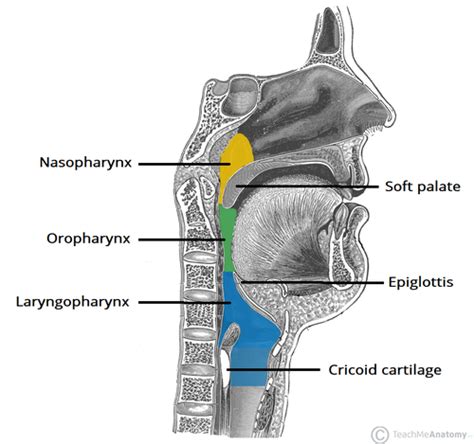
Table of Contents
The Esophagus: The Straight Tube That Transports Food from the Pharynx
The human digestive system is a marvel of biological engineering, a complex network of organs working in concert to break down food and absorb its nutrients. At the heart of this system lies a seemingly simple yet crucial component: the esophagus. Often overlooked, the esophagus plays a vital role in the process of digestion, acting as the conduit that transports food from the pharynx (throat) to the stomach. This article delves into the intricacies of the esophagus, exploring its anatomy, physiology, and potential problems, all while aiming for a comprehensive understanding of its function as the straight tube passing food from the pharynx.
Anatomy of the Esophagus: A Detailed Look
The esophagus, a muscular tube approximately 25-30 centimeters long, begins at the inferior end of the pharynx, just behind the larynx (voice box). It descends through the neck, pierces the diaphragm (the muscle separating the chest cavity from the abdomen), and finally connects to the stomach at the gastroesophageal junction. This seemingly simple structure, however, is composed of several distinct layers, each contributing to its overall function:
1. Mucosa: The Protective Inner Lining
The innermost layer, the mucosa, is a delicate mucous membrane responsible for protecting the esophageal lining from the abrasive effects of food. It's composed of stratified squamous epithelium, a type of tissue designed to withstand friction and chemical irritation. The mucus secreted by this layer lubricates the passage of food, preventing damage to the esophageal wall.
2. Submucosa: A Supportive Layer
Beneath the mucosa lies the submucosa, a layer of connective tissue rich in blood vessels, lymphatic vessels, and nerves. These vessels supply the esophagus with nutrients and oxygen, while the nerves help regulate its motility and secretions. The submucosa also houses esophageal glands, which secrete mucus to further lubricate the esophageal lumen (the internal space of the esophagus).
3. Muscularis Externa: The Engine of Propulsion
The muscularis externa is the thickest layer of the esophageal wall and is responsible for the propulsion of food towards the stomach. It comprises two layers of smooth muscle: an inner circular layer and an outer longitudinal layer. These muscles work in a coordinated fashion, creating peristaltic waves—rhythmic contractions that push the bolus (the mass of chewed food) down the esophagus. The upper third of the esophagus also contains skeletal muscle, allowing for voluntary control of swallowing.
4. Adventitia: The Outermost Layer
The outermost layer of the esophagus, the adventitia, is a connective tissue layer that anchors the esophagus to surrounding structures. It provides structural support and protection, ensuring the esophagus remains in its proper position within the thorax.
Physiology of Swallowing: A Coordinated Effort
The act of swallowing, or deglutition, is a complex process involving the coordinated action of multiple muscles and nerves. It's divided into three phases:
1. Oral Phase: Voluntary Control
The oral phase begins with the voluntary act of chewing and forming the bolus. The tongue then pushes the bolus posteriorly into the pharynx, initiating the involuntary phases of swallowing.
2. Pharyngeal Phase: Involuntary Reflex
This phase is a rapid, involuntary reflex. The soft palate elevates to prevent food from entering the nasal cavity, while the epiglottis covers the larynx to protect the airways. The pharyngeal constrictors, muscles in the pharynx, contract sequentially, propelling the bolus into the esophagus.
3. Esophageal Phase: Peristalsis Takes Over
Once the bolus enters the esophagus, the esophageal phase begins. Peristaltic waves, generated by the coordinated contractions of the circular and longitudinal muscle layers, propel the bolus toward the stomach. This process is largely involuntary, ensuring efficient transport of food even while lying down or upside down. The lower esophageal sphincter (LES), a ring of muscle at the gastroesophageal junction, relaxes to allow the bolus to enter the stomach and then contracts to prevent stomach acid from refluxing back into the esophagus.
Clinical Significance: Disorders of the Esophagus
While generally a robust structure, the esophagus can be susceptible to various disorders, some of which can severely impact its function and overall health. These include:
1. Gastroesophageal Reflux Disease (GERD): Acid Reflux
GERD is a common condition characterized by the reflux of stomach acid into the esophagus. This can cause heartburn, chest pain, and esophageal damage. The weakened LES is often a contributing factor.
2. Esophageal Cancer: A Serious Threat
Esophageal cancer is a relatively rare but serious malignancy. Risk factors include smoking, alcohol consumption, and certain dietary habits. Early detection is crucial for successful treatment.
3. Achalasia: Impaired Esophageal Motility
Achalasia is a disorder affecting the esophageal muscles. The LES fails to relax properly, hindering the passage of food into the stomach. This can lead to dysphagia (difficulty swallowing) and food accumulation in the esophagus.
4. Esophageal Diverticula: Pouches in the Esophageal Wall
Esophageal diverticula are outpouchings of the esophageal wall. These pouches can trap food, leading to infection and inflammation.
5. Mallory-Weiss Tear: Esophageal Laceration
Mallory-Weiss tears are lacerations (tears) in the esophageal lining, often caused by severe vomiting. These tears can lead to bleeding and require medical attention.
6. Esophageal Varices: Enlarged Esophageal Veins
Esophageal varices are abnormally enlarged veins in the esophagus, commonly associated with liver cirrhosis. These varices can rupture, causing life-threatening bleeding.
Diagnostic Procedures for Esophageal Issues
Various diagnostic techniques are used to evaluate esophageal function and identify potential problems. These include:
- Upper endoscopy: A procedure involving inserting a thin, flexible tube with a camera to visualize the esophageal lining.
- Esophageal manometry: Measures the pressure within the esophagus to assess its motility.
- Barium swallow: An X-ray study using barium contrast to visualize the esophagus and detect structural abnormalities.
- pH monitoring: Measures the acidity within the esophagus to diagnose GERD.
Conclusion: The Unsung Hero of Digestion
The esophagus, often overlooked in discussions of the digestive system, plays a critical role in transporting food from the pharynx to the stomach. Its intricate anatomy and precisely coordinated physiology ensure efficient and safe passage of food, preventing aspiration into the lungs and facilitating the subsequent stages of digestion. Understanding the structure and function of this seemingly simple tube, along with its associated disorders and diagnostic techniques, is crucial for maintaining overall digestive health. Further research continues to unravel the complexities of esophageal function and refine treatments for associated ailments. Continued awareness of potential issues and proactive healthcare are vital to ensuring the continued smooth operation of this unsung hero of digestion.
Latest Posts
Latest Posts
-
Hesi A2 Anatomy And Physiology Quizlet 2023
Mar 31, 2025
-
A Disorder Caused By Hyperthyroidism Is Quizlet
Mar 31, 2025
-
An Example Of An Automatic Stabilizer Is Quizlet
Mar 31, 2025
-
Budgeting For Life After High School Worksheet Answers Quizlet
Mar 31, 2025
-
An Enlargement Of The Thyroid Gland Is Called Quizlet
Mar 31, 2025
Related Post
Thank you for visiting our website which covers about A Straight Tube That Passes Food From The Pharynx Quizlet . We hope the information provided has been useful to you. Feel free to contact us if you have any questions or need further assistance. See you next time and don't miss to bookmark.
