Correctly Label The Components Of The Lungs
Breaking News Today
Mar 16, 2025 · 6 min read
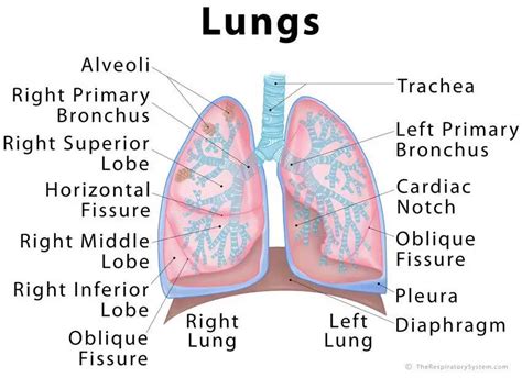
Table of Contents
Correctly Labeling the Components of the Lungs: A Comprehensive Guide
The lungs, the vital organs of respiration, are complex structures with numerous components working in concert to facilitate gas exchange. Understanding the anatomy of the lungs is crucial for anyone studying respiratory physiology, medicine, or related fields. This comprehensive guide will walk you through the correct labeling of the key components of the lungs, ensuring a thorough grasp of their intricate structure. We'll explore everything from the macroscopic lobes and fissures to the microscopic alveoli and their supporting structures.
The Gross Anatomy of the Lungs: External Features
Let's begin by examining the external features visible to the naked eye. The lungs, situated within the thoracic cavity, are paired organs—a right lung and a left lung. However, they are not perfectly symmetrical.
1. Right Lung: Larger and Divided
The right lung is larger and wider than the left lung, accommodating the position of the heart. It is divided into three lobes:
- Superior Lobe: The uppermost lobe.
- Middle Lobe: Located between the superior and inferior lobes.
- Inferior Lobe: The largest lobe, situated at the bottom.
These lobes are separated by deep fissures:
- Oblique Fissure: Separates the inferior lobe from both the superior and middle lobes.
- Horizontal Fissure: Separates the superior and middle lobes.
These fissures are crucial landmarks for clinicians during physical examination and imaging studies. Identifying them correctly is essential for accurate diagnosis.
2. Left Lung: Smaller and Two Lobed
The left lung is smaller than the right lung due to the space occupied by the heart. It is divided into two lobes:
- Superior Lobe: The upper lobe, often exhibiting a characteristic cardiac notch—a concavity accommodating the heart.
- Inferior Lobe: The lower lobe, similar in function and structure to the inferior lobe of the right lung.
Only one major fissure, the oblique fissure, separates the superior and inferior lobes of the left lung. The absence of a middle lobe is a key distinguishing characteristic between the left and right lungs.
The Bronchial Tree: The Pathway for Air
The lungs receive air through a complex branching system known as the bronchial tree. This begins with the trachea, which branches into the right and left main bronchi.
1. Main Bronchi (Primary Bronchi): Initial Branches
The right and left main bronchi are the first branches of the trachea, entering each lung at the hilum—the region where blood vessels, nerves, and lymphatics enter and exit the lungs. The right main bronchus is wider, shorter, and more vertical than the left, making it more susceptible to aspiration of foreign objects.
2. Lobar Bronchi (Secondary Bronchi): Branching into Lobes
The main bronchi further subdivide into lobar bronchi, with three on the right (one for each lobe) and two on the left (one for each lobe). These bronchi supply air to the individual lobes of the lungs.
3. Segmental Bronchi (Tertiary Bronchi): Supplying Bronchopulmonary Segments
The lobar bronchi continue to branch into segmental bronchi, supplying air to specific anatomical units called bronchopulmonary segments. These segments are functionally independent units, facilitating surgical resection if necessary. Precise identification of these segments is critical in thoracic surgery.
4. Bronchioles: Smaller Airways
The branching continues with smaller and smaller airways called bronchioles. These airways lack cartilage, unlike the larger bronchi, and are primarily composed of smooth muscle. The smooth muscle's contraction and relaxation regulate airflow, influencing the overall lung capacity and resistance.
5. Terminal Bronchioles: The End of the Conducting Zone
The bronchioles eventually lead to terminal bronchioles, marking the end of the conducting zone—the portion of the airway responsible for transporting air to the respiratory zone.
The Respiratory Zone: Where Gas Exchange Happens
The respiratory zone is where gas exchange—the crucial process of oxygen uptake and carbon dioxide elimination—occurs. The key players here are:
1. Respiratory Bronchioles: Transition Zone
Respiratory bronchioles are the transition zone between the conducting and respiratory zones. They have alveoli budding from their walls, beginning the gas exchange process.
2. Alveolar Ducts: Connecting Airways
Alveolar ducts are small airways that lead from the respiratory bronchioles to the alveolar sacs. They are primarily composed of alveoli.
3. Alveolar Sacs: Grape-like Clusters
Alveolar sacs are grape-like clusters of alveoli, providing a vast surface area for gas exchange. Their structure maximizes the efficiency of oxygen and carbon dioxide diffusion.
4. Alveoli: The Functional Units of Gas Exchange
Alveoli are tiny, thin-walled air sacs, the primary sites of gas exchange. Their thin walls are composed of a single layer of epithelial cells (type I pneumocytes) facilitating efficient diffusion of gases. Within their walls are also type II pneumocytes responsible for producing surfactant, a crucial substance that reduces surface tension in the alveoli, preventing collapse.
Supporting Structures: Essential for Lung Function
The proper functioning of the lungs relies on various supporting structures:
1. Pulmonary Arteries and Veins: Blood Supply
The pulmonary arteries carry deoxygenated blood from the heart to the lungs for oxygenation. The pulmonary veins then return the oxygenated blood to the heart. This circulatory system is unique in that it carries blood low in oxygen to the lungs and returns oxygenated blood to the heart.
2. Bronchial Arteries and Veins: Nutritional Supply
The bronchial arteries supply oxygenated blood to the lung tissues themselves, providing nourishment for the bronchi, bronchioles, and other lung structures. The bronchial veins drain this blood.
3. Pleura: Protective Membranes
The lungs are enveloped by a double-layered membrane called the pleura. The visceral pleura is closely adhered to the lung surface, while the parietal pleura lines the thoracic cavity. The space between these layers, the pleural cavity, contains a small amount of pleural fluid that lubricates the surfaces and helps to maintain lung expansion.
4. Lymphatic System: Drainage and Defense
The lymphatic system within the lungs plays a crucial role in draining excess fluid and removing waste products. It also plays a significant role in the immune defense of the lungs. The lymph nodes in the lungs filter harmful substances and contribute to the overall defense against infections.
5. Nerves: Innervation and Control
The lungs receive innervation from both the sympathetic and parasympathetic nervous systems. This allows for the regulation of bronchiole diameter, influencing airflow and overall respiratory function.
Clinical Significance of Accurate Labeling
Accurate labeling of lung components is crucial in various clinical settings. For example:
- Radiology: Precise identification of lobes, fissures, and bronchopulmonary segments on chest X-rays and CT scans is vital for diagnosing and managing lung diseases.
- Thoracic Surgery: Surgeons rely on a thorough understanding of lung anatomy to plan and perform procedures like lobectomy (surgical removal of a lobe) or segmentectomy (surgical removal of a segment).
- Pulmonology: Respiratory specialists need a detailed knowledge of lung anatomy to diagnose and treat respiratory conditions such as pneumonia, bronchitis, asthma, and lung cancer.
Conclusion: Mastering Lung Anatomy
Correctly labeling the components of the lungs is essential for understanding their function and for effectively diagnosing and treating lung diseases. This detailed guide provides a comprehensive overview of the lung's intricate structure, from the macroscopic lobes and fissures to the microscopic alveoli. By mastering this knowledge, you’ll gain a deeper understanding of the respiratory system and its critical role in maintaining overall health. Remember, constant review and visualization are key to retaining this information effectively. Consider utilizing anatomical models, interactive diagrams, and medical imaging examples to reinforce your understanding. This multifaceted approach will significantly improve your ability to accurately label and comprehend the complex anatomy of the lungs.
Latest Posts
Latest Posts
-
According To Stalin What Must Soviets Do To Defeat Hitler
Mar 16, 2025
-
Cpr Test Questions And Answers American Heart Association
Mar 16, 2025
-
What Hostile Intelligence Collection Method Is The Process Of Obtaining
Mar 16, 2025
-
State Of Tennessee F Endorsement Practice Test
Mar 16, 2025
-
Pogil Control Of Gene Expression In Prokaryotes
Mar 16, 2025
Related Post
Thank you for visiting our website which covers about Correctly Label The Components Of The Lungs . We hope the information provided has been useful to you. Feel free to contact us if you have any questions or need further assistance. See you next time and don't miss to bookmark.
