Correctly Label The Following Anatomical Features Of The Spinal Cord
Breaking News Today
Mar 25, 2025 · 6 min read
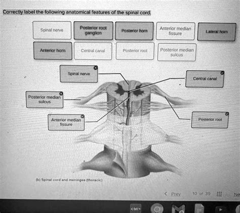
Table of Contents
Correctly Labeling the Anatomical Features of the Spinal Cord: A Comprehensive Guide
The spinal cord, a crucial part of the central nervous system, is a complex structure responsible for transmitting signals between the brain and the rest of the body. Understanding its intricate anatomy is fundamental to comprehending neurological function and dysfunction. This comprehensive guide will delve into the key anatomical features of the spinal cord, providing detailed descriptions and aiding in their accurate labeling. We'll explore both macroscopic and microscopic features, equipping you with a solid understanding of this vital organ.
Macroscopic Anatomy of the Spinal Cord: External Features
The spinal cord, roughly cylindrical in shape, extends from the medulla oblongata of the brainstem to approximately the level of the first lumbar vertebra (L1). Its external features are readily identifiable and crucial for understanding its overall structure.
1. Spinal Cord Length and Segments:
The spinal cord isn't a continuous, uniform structure. Instead, it's segmented, with each segment giving rise to a pair of spinal nerves. These segments are crucial for understanding the organization of the spinal cord's function. There are 31 pairs of spinal nerves:
- 8 Cervical (C1-C8): Supply the neck, shoulders, arms, and hands.
- 12 Thoracic (T1-T12): Innervate the chest, abdomen, and back.
- 5 Lumbar (L1-L5): Supply the lower back, hips, and front of the legs.
- 5 Sacral (S1-S5): Innervate the buttocks, genitalia, and back of the legs.
- 1 Coccygeal (Co1): Innervates a small area of the skin over the coccyx.
It's important to note that the spinal cord's length is shorter than the vertebral column. The spinal nerves continue beyond the end of the spinal cord as the cauda equina, a collection of nerve roots resembling a horse's tail.
2. Spinal Cord Enlargements:
Two noticeable enlargements mark the spinal cord:
- Cervical Enlargement (C4-T1): This thicker region provides the neural pathways for innervation of the upper limbs. The increased size reflects the greater number of neurons required to control the complex movements of the arms and hands.
- Lumbosacral Enlargement (L1-S3): Similarly, this enlargement supports the innervation of the lower limbs. The larger size reflects the neural demands of leg and foot movement.
These enlargements are critical for understanding the distribution of motor and sensory neurons within the spinal cord.
3. Conus Medullaris and Filum Terminale:
The spinal cord tapers to a cone-shaped structure known as the conus medullaris at the level of L1-L2. Extending from the conus medullaris is the filum terminale, a slender, fibrous strand that anchors the spinal cord to the coccyx, providing structural support. The filum terminale is crucial for maintaining the spinal cord's position within the vertebral column.
4. Anterior Median Fissure and Posterior Median Sulcus:
The spinal cord's surface is marked by two prominent grooves:
- Anterior Median Fissure: A deep, longitudinal groove on the anterior (front) surface of the spinal cord.
- Posterior Median Sulcus: A shallower, longitudinal groove on the posterior (back) surface of the spinal cord.
These grooves are important landmarks that help to divide the spinal cord into right and left halves.
5. Spinal Nerve Roots:
Each spinal segment gives rise to a pair of spinal nerves, which emerge from the spinal cord through the intervertebral foramina. Each spinal nerve has two roots:
- Anterior (Ventral) Root: Contains motor (efferent) fibers carrying signals away from the central nervous system to muscles and glands.
- Posterior (Dorsal) Root: Contains sensory (afferent) fibers carrying signals towards the central nervous system from sensory receptors in the periphery. Each posterior root also has a dorsal root ganglion, containing the cell bodies of sensory neurons.
Microscopic Anatomy of the Spinal Cord: Internal Features
Moving beyond the external features, we now explore the intricate internal structure of the spinal cord. Understanding its internal organization is critical for comprehending how information is processed and transmitted.
1. Gray Matter and White Matter:
In cross-section, the spinal cord reveals a characteristic "H" or butterfly shape of gray matter surrounded by white matter.
-
Gray Matter: Primarily composed of neuronal cell bodies, dendrites, and unmyelinated axons. It's involved in processing information and initiating reflexes. The gray matter is organized into horns:
- Anterior (Ventral) Horns: Contain motor neuron cell bodies that innervate skeletal muscles.
- Posterior (Dorsal) Horns: Receive sensory information from the body.
- Lateral Horns: Present only in the thoracic and upper lumbar regions, containing the cell bodies of sympathetic preganglionic neurons involved in the autonomic nervous system.
-
White Matter: Composed largely of myelinated axons organized into ascending and descending tracts. These tracts facilitate communication between different levels of the spinal cord and the brain. The white matter is further divided into columns or funiculi:
- Posterior (Dorsal) Columns: Carry sensory information, such as touch, pressure, and proprioception (body position).
- Lateral Columns: Contain both ascending and descending tracts, including those involved in pain, temperature, and motor control.
- Anterior (Ventral) Columns: Carry both ascending and descending tracts, including motor information.
2. Ascending and Descending Tracts:
The white matter tracts are crucial for the spinal cord's function in transmitting information between the brain and the periphery. These tracts are often named according to their origin and termination points, and their specific functions. Examples include:
- Ascending Tracts (Sensory): Dorsal Column-Medial Lemniscus pathway (touch, pressure, proprioception); Spinothalamic tract (pain, temperature); Spinocerebellar tracts (proprioception).
- Descending Tracts (Motor): Corticospinal tract (voluntary movement); Rubrospinal tract (muscle tone); Reticulospinal tract (posture and balance); Vestibulospinal tract (balance and posture).
3. Central Canal:
The center of the gray matter contains the central canal, a small, fluid-filled cavity that is continuous with the ventricles of the brain. This canal is lined with ependymal cells and is filled with cerebrospinal fluid (CSF). The CSF provides cushioning and nutrient delivery to the spinal cord.
Clinical Significance of Spinal Cord Anatomy
Understanding the spinal cord's anatomy is crucial for diagnosing and treating various neurological conditions. Damage to the spinal cord, such as from injury or disease, can lead to a range of impairments depending on the location and extent of the damage. For example:
- Spinal Cord Injury: Trauma can sever or compress the spinal cord, leading to loss of motor and sensory function below the level of the injury. The specific deficits depend on the location and severity of the damage.
- Multiple Sclerosis (MS): An autoimmune disease that attacks the myelin sheath surrounding axons in the central nervous system, leading to a range of neurological symptoms, including weakness, numbness, and vision problems.
- Amyotrophic Lateral Sclerosis (ALS): A progressive neurodegenerative disease that affects motor neurons, leading to muscle weakness and atrophy.
Conclusion: Accurate Labeling and Comprehensive Understanding
Correctly labeling the anatomical features of the spinal cord requires a thorough understanding of its macroscopic and microscopic structure. From the external landmarks such as the anterior median fissure and posterior median sulcus to the internal organization of gray and white matter, and the specific functions of the various tracts, accurate identification is essential for grasping the complexity of the spinal cord's function. This detailed overview provides a robust framework for comprehending the intricacies of the spinal cord and its role in the nervous system. Remember, this knowledge is not only crucial for medical professionals but also essential for anyone seeking a deeper understanding of human anatomy and physiology. Continued study and review will further solidify this knowledge base.
Latest Posts
Latest Posts
-
Hourly Retail Associate Assessment Walmart Answers 2024
Mar 28, 2025
-
Traditional Savings Account Typical Add To Balance Regularly
Mar 28, 2025
-
Costs That Can Be Traced Directly To A Segment
Mar 28, 2025
-
Blank Refers To The Soil Removed From An Excavation
Mar 28, 2025
-
How Can Producers Maximize Their Profit Check All That Apply
Mar 28, 2025
Related Post
Thank you for visiting our website which covers about Correctly Label The Following Anatomical Features Of The Spinal Cord . We hope the information provided has been useful to you. Feel free to contact us if you have any questions or need further assistance. See you next time and don't miss to bookmark.
