Correctly Label The Following Coronary Blood Vessels Of The Heart
Breaking News Today
Mar 28, 2025 · 6 min read
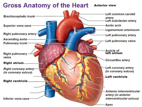
Table of Contents
Correctly Labeling the Coronary Blood Vessels of the Heart: A Comprehensive Guide
The heart, the tireless engine of our bodies, demands a robust and efficient blood supply to function optimally. This supply is provided by the coronary arteries, a network of vessels that branch across the heart's surface, delivering oxygen-rich blood to the cardiac muscle. Understanding the intricate anatomy of these vessels is crucial for healthcare professionals, medical students, and anyone interested in cardiovascular health. This comprehensive guide will delve into the correct labeling of the major coronary blood vessels, emphasizing their branching patterns and clinical significance.
The Major Coronary Arteries: Origin and Distribution
The coronary arteries originate from the aorta, the body's largest artery, just beyond the aortic valve. These arteries, along with their branches, form a complex network ensuring every part of the heart muscle receives sufficient blood flow. Two main arteries dominate this network:
1. The Right Coronary Artery (RCA)
The RCA typically arises from the right aortic sinus, just above the aortic valve. Its course and branching pattern can vary significantly between individuals, but generally, it travels along the atrioventricular groove (the groove between the atria and ventricles) on the right side of the heart.
Key Branches of the RCA:
- Sinus Node Artery (SA Node Artery): This crucial branch supplies blood to the sinoatrial node, the heart's natural pacemaker, responsible for initiating the heartbeat. Its origin is variable; in approximately 60% of individuals, it arises from the RCA.
- Right Atrial Branches: These branches supply blood to the right atrium.
- Right Ventricular Branches: These branches supply blood to the right ventricle.
- Posterior Descending Artery (PDA): This is a significant branch of the RCA in approximately 85% of individuals. It runs down the posterior interventricular groove, supplying blood to the posterior walls of both ventricles and the posterior interventricular septum. The PDA's dominance is clinically important as it indicates the primary blood supply to the posterior heart.
- Acute Marginal Branches: These branches supply blood to the right ventricle.
2. The Left Coronary Artery (LCA)
The LCA, usually larger than the RCA, originates from the left aortic sinus, also just above the aortic valve. It quickly divides into two major branches:
Key Branches of the LCA:
- Left Anterior Descending Artery (LAD): Also known as the anterior interventricular artery, the LAD is the largest branch of the LCA. It descends along the anterior interventricular groove, supplying blood to the anterior walls of both ventricles and the anterior two-thirds of the interventricular septum. The LAD is often described as the "widow maker" due to its critical role in supplying a large portion of the heart muscle; occlusion here can lead to a significant myocardial infarction (heart attack).
- Circumflex Artery (Cx): The Cx artery curves around the left side of the heart in the atrioventricular groove. It supplies blood to the left atrium and the lateral and posterior walls of the left ventricle. It also frequently gives rise to the left marginal branch, supplying blood to the left ventricle.
Variations in Coronary Artery Anatomy
It's crucial to remember that the coronary artery anatomy is highly variable. While the general pattern described above is common, significant variations exist, impacting both the clinical presentation of coronary artery disease (CAD) and interventional strategies. Some key variations include:
- Dominance: The term "dominance" refers to which coronary artery supplies the posterior descending artery (PDA). Right dominance, the most common pattern (approximately 85% of individuals), means the RCA gives rise to the PDA. Left dominance (approximately 8% of individuals) means the circumflex artery supplies the PDA. Co-dominance (approximately 7% of individuals) occurs when both the RCA and the Cx contribute to the PDA. Understanding dominance is crucial for interpreting coronary angiograms and predicting the impact of arterial blockage.
- Origin and Course: The origin and course of the coronary arteries, and their branches, can vary considerably. Some individuals may have additional or unusual branches, while others may have smaller or absent branches. These variations can influence the clinical presentation of coronary artery disease.
- Anastomoses: Anastomoses are connections between different branches of the coronary arteries. These connections provide alternative pathways for blood flow, which can be vital in situations where one artery is blocked or narrowed. The extent and distribution of these anastomoses are variable and affect the vulnerability of the myocardium to ischemia (lack of oxygen).
Clinical Significance of Coronary Artery Anatomy
Knowledge of coronary artery anatomy is paramount in diagnosing and treating cardiovascular diseases.
- Coronary Artery Disease (CAD): CAD is characterized by the buildup of plaque (atherosclerosis) within the coronary arteries, reducing blood flow to the heart muscle. Understanding the precise location and extent of the blockage is crucial for determining the best treatment strategy. Angiography, a procedure where a dye is injected into the coronary arteries to visualize them via X-ray, is essential for identifying narrowed or blocked arteries.
- Myocardial Infarction (Heart Attack): A heart attack occurs when blood flow to a portion of the heart muscle is abruptly cut off, usually due to a complete blockage of a coronary artery. The location of the blockage determines which part of the heart is affected and the severity of the heart attack.
- Coronary Artery Bypass Grafting (CABG): CABG surgery involves creating new pathways for blood to flow around blocked coronary arteries. A surgeon will carefully plan the bypass grafts based on a thorough understanding of the patient's coronary artery anatomy.
- Percutaneous Coronary Intervention (PCI): PCI, also known as angioplasty, involves inserting a catheter into a blocked coronary artery to open it up and restore blood flow. Knowledge of coronary artery anatomy guides the placement of the catheter and the deployment of stents.
Labeling the Coronary Arteries: A Practical Approach
Accurate labeling of the coronary arteries requires careful observation and understanding of their branching patterns and spatial relationships. Here's a structured approach:
- Identify the Aorta: Begin by identifying the aorta, the large artery arising from the heart.
- Locate the Coronary Ostia: The coronary arteries originate from the aorta just beyond the aortic valve. Identify the right and left coronary ostia (openings).
- Trace the RCA: Follow the course of the RCA along the atrioventricular groove on the right side of the heart. Identify its major branches: the sinus node artery (if present), right atrial branches, right ventricular branches, and the posterior descending artery (if present).
- Trace the LCA: Follow the course of the LCA, noting its bifurcation into the LAD and the Cx artery. Trace the LAD along the anterior interventricular groove and the Cx around the left side of the heart. Identify their respective branches.
- Determine Dominance: Based on the origin of the PDA, determine whether the heart exhibits right dominance, left dominance, or co-dominance.
- Note Variations: Be aware that variations in coronary artery anatomy are common. Document any unusual branching patterns or anatomical variations observed.
Conclusion
Correctly labeling the coronary blood vessels of the heart is a fundamental skill for understanding cardiovascular anatomy and physiology. This guide has provided a detailed overview of the major coronary arteries, their branches, variations in anatomy, and their clinical significance. By mastering this knowledge, healthcare professionals can better diagnose and treat cardiovascular diseases, ultimately improving patient outcomes. Further study and practical experience, such as observing anatomical models and coronary angiograms, are crucial for solidifying this crucial understanding. Remember to always consult reliable anatomical resources and medical textbooks for comprehensive and up-to-date information.
Latest Posts
Latest Posts
-
The Best Temperature For Short Term Refrigeration Storage Is
Mar 31, 2025
-
The Demand Curve Can Only Shift In One Direction
Mar 31, 2025
-
Explain The Role That Heredity Plays In Skill Related Fitness
Mar 31, 2025
-
A Main Purpose Of This Rbt Training Is To Quizlet
Mar 31, 2025
-
Common Causes Of Syncope In Older Patients Quizlet
Mar 31, 2025
Related Post
Thank you for visiting our website which covers about Correctly Label The Following Coronary Blood Vessels Of The Heart . We hope the information provided has been useful to you. Feel free to contact us if you have any questions or need further assistance. See you next time and don't miss to bookmark.
