Correctly Label The Following Internal Anatomy Of The Heart
Breaking News Today
Mar 19, 2025 · 6 min read
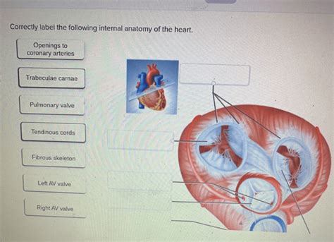
Table of Contents
Correctly Labeling the Internal Anatomy of the Heart: A Comprehensive Guide
The human heart, a remarkable organ, tirelessly pumps blood throughout the body, sustaining life itself. Understanding its intricate internal anatomy is crucial for anyone studying medicine, biology, or simply curious about the mechanics of this vital organ. This comprehensive guide will delve into the detailed structure of the heart, providing clear explanations and visual aids to help you correctly label its internal components. We’ll explore the chambers, valves, vessels, and conducting system, clarifying their roles and interrelationships.
The Four Chambers: The Heart's Pumping Powerhouses
The heart is divided into four chambers: two atria (singular: atrium) and two ventricles. These chambers work in a coordinated sequence to ensure efficient blood circulation.
1. The Right Atrium:
The right atrium receives deoxygenated blood returning from the body via the superior vena cava (carrying blood from the upper body) and the inferior vena cava (carrying blood from the lower body). It also receives blood from the coronary sinus, which drains blood from the heart muscle itself. The right atrium's muscular wall is relatively thin compared to the ventricles because it doesn't need to generate as much force to pump blood. The tricuspid valve, a crucial component, prevents the backflow of blood into the right atrium when the right ventricle contracts.
2. The Right Ventricle:
The right ventricle receives deoxygenated blood from the right atrium and pumps it to the lungs via the pulmonary artery. The pulmonary artery is unique as it's the only artery in the body carrying deoxygenated blood. The right ventricle has a thicker muscular wall than the right atrium, reflecting its need to generate enough pressure to push blood through the pulmonary circulation. The pulmonary valve prevents the backflow of blood from the pulmonary artery into the right ventricle.
3. The Left Atrium:
The left atrium receives oxygenated blood from the lungs through the pulmonary veins. These are the only veins in the body carrying oxygenated blood. The left atrium's relatively thin muscular wall facilitates the passive transfer of blood to the left ventricle.
4. The Left Ventricle:
The left ventricle is the powerhouse of the heart. It receives oxygenated blood from the left atrium and pumps it to the rest of the body via the aorta. The left ventricle has the thickest muscular wall of all the chambers, reflecting the high pressure required to propel blood throughout the systemic circulation. The mitral valve (also known as the bicuspid valve), prevents backflow into the left atrium during ventricular contraction. The aortic valve prevents backflow from the aorta into the left ventricle.
The Heart Valves: Ensuring One-Way Blood Flow
The heart valves are crucial for maintaining unidirectional blood flow. Their precise opening and closing prevent backflow, ensuring efficient circulation.
1. Atrioventricular (AV) Valves:
- Tricuspid Valve: Located between the right atrium and right ventricle. It has three cusps (leaflets) and prevents backflow from the ventricle to the atrium.
- Mitral Valve (Bicuspid Valve): Located between the left atrium and left ventricle. It has two cusps and prevents backflow from the ventricle to the atrium.
2. Semilunar Valves:
- Pulmonary Valve: Located between the right ventricle and the pulmonary artery. It prevents backflow from the pulmonary artery to the right ventricle.
- Aortic Valve: Located between the left ventricle and the aorta. It prevents backflow from the aorta to the left ventricle.
The coordinated opening and closing of these valves are essential for the efficient pumping action of the heart. Improper valve function can lead to various heart conditions.
The Heart's Electrical Conduction System: The Pacemaker and More
The heart's electrical conduction system is a complex network that regulates the rhythmic contraction of the heart muscle. This system ensures the coordinated pumping action of the atria and ventricles.
1. Sinoatrial (SA) Node (the Pacemaker):
Located in the right atrium, the SA node generates electrical impulses that initiate the heartbeat. It's the natural pacemaker of the heart, setting the rhythm for the rest of the conduction system.
2. Atrioventricular (AV) Node:
Located in the interatrial septum, the AV node receives impulses from the SA node and delays their transmission to the ventricles. This delay ensures that the atria contract before the ventricles, allowing for efficient blood filling.
3. Bundle of His:
This specialized bundle of fibers conducts impulses from the AV node to the ventricles. It divides into the right and left bundle branches.
4. Purkinje Fibers:
These fibers spread throughout the ventricular walls, rapidly distributing the electrical impulses to ensure coordinated contraction of the ventricles.
Major Blood Vessels Connected to the Heart
Several major blood vessels connect to the heart, facilitating the continuous flow of blood.
1. Superior and Inferior Vena Cavae:
These large veins return deoxygenated blood from the systemic circulation to the right atrium.
2. Pulmonary Artery:
This artery carries deoxygenated blood from the right ventricle to the lungs for oxygenation.
3. Pulmonary Veins:
These veins return oxygenated blood from the lungs to the left atrium.
4. Aorta:
This major artery carries oxygenated blood from the left ventricle to the rest of the body.
5. Coronary Arteries:
These arteries branch off the aorta and supply oxygenated blood to the heart muscle itself. Blockages in the coronary arteries can lead to heart attacks.
Understanding the Cardiac Cycle: A Coordinated Dance
The cardiac cycle refers to the sequence of events that occur during one complete heartbeat. This involves the coordinated contraction and relaxation of the atria and ventricles, facilitated by the electrical conduction system and the precise functioning of the heart valves.
Diastole (Relaxation):
During diastole, the heart muscle relaxes, allowing the chambers to fill with blood. The atria fill passively, and then actively contract, pushing blood into the ventricles.
Systole (Contraction):
During systole, the ventricles contract, forcefully pumping blood into the pulmonary artery (right ventricle) and the aorta (left ventricle). The atrioventricular valves close to prevent backflow into the atria, while the semilunar valves open to allow blood to exit the ventricles.
Clinical Significance and Related Conditions
A thorough understanding of the heart's internal anatomy is paramount in diagnosing and treating various cardiovascular conditions. Conditions affecting the chambers, valves, or conduction system can have serious consequences.
1. Valvular Heart Disease:
This involves malfunctioning heart valves, leading to either stenosis (narrowing) or regurgitation (leakage). Symptoms can range from mild shortness of breath to severe heart failure.
2. Congenital Heart Defects:
These are structural abnormalities present at birth, affecting the heart's chambers, valves, or major vessels. These defects can vary widely in severity.
3. Coronary Artery Disease (CAD):
This involves the build-up of plaque in the coronary arteries, reducing blood flow to the heart muscle and potentially leading to heart attacks.
4. Arrhythmias:
These are irregularities in the heart's rhythm, caused by disturbances in the electrical conduction system. They can range from mild palpitations to life-threatening conditions.
Conclusion: Mastering the Heart's Internal Landscape
Successfully labeling the internal anatomy of the heart requires careful study and a comprehensive understanding of the individual components and their interrelationships. This guide provides a detailed roadmap for achieving this goal, equipping you with the knowledge to appreciate the complexity and elegance of this vital organ. Remember, the heart's intricate structure is crucial for its function, and understanding this structure is fundamental to understanding its role in maintaining life itself. Continue your exploration of cardiac anatomy and physiology to gain a deeper appreciation for this fascinating and essential organ. By thoroughly understanding these structures and their function, you'll be well on your way to mastering the intricacies of the human heart.
Latest Posts
Latest Posts
-
Explain The Difference Between Physical Activity And Exercise
Mar 19, 2025
-
The Functional Unit Of The Kidney Is The
Mar 19, 2025
-
Compare And Contrast Mental Health And Emotional Health
Mar 19, 2025
-
Where Are Metals Located On The Periodic Table
Mar 19, 2025
-
What School Did Brandon Go To In Ground Zero
Mar 19, 2025
Related Post
Thank you for visiting our website which covers about Correctly Label The Following Internal Anatomy Of The Heart . We hope the information provided has been useful to you. Feel free to contact us if you have any questions or need further assistance. See you next time and don't miss to bookmark.
