Correctly Label The Following Parts Of The Digestive System
Breaking News Today
Mar 30, 2025 · 7 min read
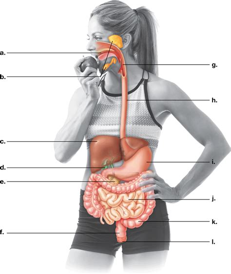
Table of Contents
Correctly Label the Following Parts of the Digestive System: A Comprehensive Guide
The digestive system is a marvel of biological engineering, a complex network responsible for breaking down the food we eat into usable nutrients. Understanding its intricate components is key to appreciating its functionality and maintaining optimal health. This comprehensive guide will explore each part of the digestive system, providing detailed descriptions and emphasizing correct labeling. We'll cover everything from the initial intake of food to the elimination of waste products.
The Oral Cavity: Where Digestion Begins
The journey of digestion starts in the oral cavity, also known as the mouth. This is where the mechanical and chemical processes of digestion first begin. Let's break down the key components:
1. Lips (Labia): The Gatekeepers of Digestion
The lips (labia), fleshy folds surrounding the mouth opening, play a crucial role in food intake. They help us grasp and manipulate food, guiding it into the oral cavity. Their sensitivity allows us to assess the texture and temperature of food before ingestion.
2. Teeth (Dentes): The Mechanical Processors
Teeth (dentes) are essential for the mechanical breakdown of food. Different types of teeth—incisors, canines, premolars, and molars—are specialized for various functions, from cutting and tearing to grinding and crushing. This process increases the surface area of food, making it easier for enzymes to act upon it. Proper dental hygiene is crucial for maintaining healthy teeth, which are vital for efficient digestion.
3. Tongue (Lingua): The Taste Sensor and Food Manipulator
The tongue (lingua) is a muscular organ covered in taste buds, responsible for taste perception and food manipulation. Its movements help mix food with saliva, forming a bolus, a soft, easily swallowed mass. The tongue also plays a critical role in swallowing, pushing the bolus towards the pharynx.
4. Salivary Glands: Initiating Chemical Digestion
Salivary glands secrete saliva, a watery fluid containing enzymes like amylase. Amylase begins the chemical breakdown of carbohydrates, initiating the process of digestion even before food reaches the stomach. Saliva also lubricates the food bolus, making it easier to swallow. Three major pairs of salivary glands exist: parotid, submandibular, and sublingual glands. Each contributes to the overall composition and volume of saliva.
5. Palatine Tonsils: The First Line of Immune Defense
Located at the back of the throat, the palatine tonsils are part of the body's lymphatic system and play an important role in immune defense. They help trap pathogens entering the body through the mouth and initiate an immune response. While their role in digestion is indirect, their health is crucial for overall digestive well-being.
The Pharynx and Esophagus: Transportation to the Stomach
After mastication (chewing) in the oral cavity, the food bolus moves into the pharynx, a passageway shared by both the respiratory and digestive systems. The epiglottis, a flap of cartilage, covers the trachea (windpipe) during swallowing, preventing food from entering the lungs.
From the pharynx, the bolus enters the esophagus, a muscular tube that transports food to the stomach through peristalsis. Peristalsis is a series of coordinated muscle contractions that propel the bolus downwards. The lower esophageal sphincter, a ring of muscle at the bottom of the esophagus, relaxes to allow food entry into the stomach and then contracts to prevent stomach acid from refluxing back into the esophagus.
The Stomach: Chemical Breakdown and Storage
The stomach is a J-shaped muscular organ that serves as a temporary storage reservoir for food. Its strong muscular walls churn and mix the food, further breaking it down mechanically. The stomach also secretes gastric juices, containing hydrochloric acid (HCl) and enzymes like pepsin.
1. Gastric Glands: Secreting Digestive Juices
Gastric glands located within the stomach lining secrete a variety of substances. Hydrochloric acid (HCl) creates a highly acidic environment, killing ingested bacteria and activating pepsinogen, an inactive form of the enzyme pepsin. Pepsin begins the digestion of proteins. Mucus secreted by goblet cells protects the stomach lining from the corrosive effects of HCl. Intrinsic factor, a glycoprotein, is essential for the absorption of vitamin B12 in the small intestine.
2. Cardiac Sphincter: Preventing Acid Reflux
The cardiac sphincter (also known as the lower esophageal sphincter) is crucial in preventing the reflux of stomach acid back into the esophagus, which can cause heartburn and other digestive issues. Its proper functioning is vital for maintaining digestive health.
3. Pyloric Sphincter: Regulating Stomach Emptying
The pyloric sphincter is a ring of muscle that controls the release of chyme (partially digested food) from the stomach into the small intestine. This controlled release ensures that the small intestine is not overwhelmed by the acidic chyme.
The Small Intestine: Nutrient Absorption
The small intestine, the longest part of the digestive tract, is responsible for the majority of nutrient absorption. It's divided into three sections: the duodenum, jejunum, and ileum.
1. Duodenum: Receiving Chyme and Pancreatic Secretions
The duodenum, the first part of the small intestine, receives chyme from the stomach, along with pancreatic juices and bile from the liver and gallbladder. These secretions further break down carbohydrates, proteins, and fats.
2. Jejunum and Ileum: Nutrient Absorption Hub
The jejunum and ileum, the middle and final sections of the small intestine, are lined with villi and microvilli, finger-like projections that significantly increase the surface area available for nutrient absorption. Nutrients are absorbed through the walls of the small intestine and transported into the bloodstream.
3. Brunner's Glands: Protecting the Duodenal Lining
Located in the submucosa of the duodenum, Brunner's glands secrete an alkaline mucus that neutralizes the acidic chyme entering from the stomach, protecting the duodenal lining from damage.
The Pancreas, Liver, and Gallbladder: Accessory Organs
Several accessory organs play vital roles in digestion, even though food doesn't directly pass through them.
1. Pancreas: Enzyme Production and Hormone Regulation
The pancreas secretes pancreatic juices containing enzymes that break down carbohydrates, proteins, and fats. It also produces hormones like insulin and glucagon, which regulate blood sugar levels.
2. Liver: Bile Production and Detoxification
The liver produces bile, a greenish-yellow fluid that emulsifies fats, breaking them down into smaller droplets for easier digestion and absorption. The liver also plays a crucial role in detoxification, filtering harmful substances from the blood.
3. Gallbladder: Bile Storage and Release
The gallbladder stores and concentrates bile produced by the liver. When needed, it releases bile into the duodenum to aid in fat digestion.
The Large Intestine: Water Absorption and Waste Elimination
The large intestine, also known as the colon, receives indigestible material from the small intestine. Its primary functions are water absorption, electrolyte absorption, and the formation and elimination of feces.
1. Cecum: The First Part of the Large Intestine
The cecum, a pouch-like structure at the beginning of the large intestine, receives waste material from the ileum. The appendix, a small, finger-like projection attached to the cecum, has a debated role in immunity.
2. Colon: Water Absorption and Waste Compaction
The colon is divided into ascending, transverse, descending, and sigmoid sections. As waste moves through the colon, water is absorbed, compacting the remaining material into feces. Bacteria residing in the colon ferment undigested carbohydrates, producing some vitamins.
3. Rectum and Anus: Waste Elimination
The rectum stores feces until elimination. When the rectum is full, the urge to defecate arises. The anus, the terminal end of the digestive tract, contains internal and external anal sphincters, which control the release of feces.
Conclusion: A Symphony of Digestion
The digestive system is a complex and interconnected network of organs working in harmony. Understanding the individual roles of each component – from the lips to the anus – is crucial for appreciating its intricate functionality and maintaining overall health. Correctly labeling these parts is fundamental to mastering the intricacies of human anatomy and physiology. Remember that this detailed guide serves as a foundation for further exploration and a deeper understanding of the fascinating world of digestion. By understanding this system, you can better appreciate the importance of a healthy diet and lifestyle in maintaining optimal digestive health.
Latest Posts
Latest Posts
-
A Patient Who Has Experienced A Back Injury
Apr 01, 2025
-
Define Back Channel Cues List 3 Examples Of Backchannel Cues
Apr 01, 2025
-
How Does Madison Use Comparison To Bolster His Argument
Apr 01, 2025
-
Afferent Signals From External Stimuli Are Carried By The
Apr 01, 2025
-
Why It Matters That Teens Are Reading Less
Apr 01, 2025
Related Post
Thank you for visiting our website which covers about Correctly Label The Following Parts Of The Digestive System . We hope the information provided has been useful to you. Feel free to contact us if you have any questions or need further assistance. See you next time and don't miss to bookmark.
