Correctly Label The Parts Of The Glomerular Filtration Membrane
Breaking News Today
Mar 20, 2025 · 6 min read
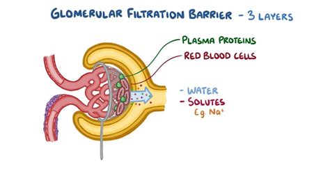
Table of Contents
Correctly Labeling the Parts of the Glomerular Filtration Membrane
The glomerular filtration membrane (GFM) is a highly specialized structure responsible for filtering blood in the kidneys. Understanding its intricate components is crucial for grasping the process of glomerular filtration and the overall function of the urinary system. This detailed guide will walk you through the precise labeling of each part of the GFM, exploring its structure and function in a clear and comprehensive manner. We'll delve into the three main layers, highlighting their individual contributions to the selective permeability of the membrane. Accurate identification of these layers is key to comprehending renal physiology and the pathogenesis of various kidney diseases.
The Three Layers of the Glomerular Filtration Membrane
The GFM isn't a single, homogeneous structure; rather, it's a tri-layered membrane composed of:
- Fenestrated Endothelium: This is the innermost layer, lining the glomerular capillaries.
- Glomerular Basement Membrane (GBM): Sandwiched between the endothelium and the podocytes, this is the crucial filtering layer.
- Podocyte Filtration Slit Diaphragm: The outermost layer, formed by the interdigitating foot processes (pedicels) of the podocytes.
Let's examine each layer in detail:
1. Fenestrated Endothelium: The Initial Barrier
The fenestrated endothelium of the glomerular capillaries forms the first layer of the GFM. The word "fenestrated" itself hints at its unique characteristic: numerous pores or fenestrae that perforate the endothelial cells. These fenestrae are approximately 70-100 nm in diameter and are significantly larger than the pores found in the endothelium of other capillaries. This larger pore size allows for relatively unrestricted passage of water and small solutes, but importantly, prevents the passage of blood cells and larger proteins.
Key Characteristics of the Fenestrated Endothelium:
- High Permeability: Due to the presence of fenestrae, the fenestrated endothelium exhibits significantly high permeability compared to other capillary beds.
- Negatively Charged Glycocaylx: The endothelial glycocalyx, a layer of glycoproteins and proteoglycans coating the endothelial surface, carries a negative charge. This negative charge further contributes to the selective permeability of the membrane, repelling negatively charged proteins and preventing their passage into the filtrate.
- Diaphragm-like structures: Although significantly larger than those in other capillaries, the fenestrae aren’t completely open. They are partially covered by a thin diaphragm that further regulates the passage of molecules.
2. Glomerular Basement Membrane (GBM): The Selective Filter
The glomerular basement membrane (GBM) is the central and arguably most important component of the GFM. This acellular structure is composed of a complex network of collagen type IV, laminin, nidogen, and heparin sulfate proteoglycans. These molecules assemble into a highly organized meshwork with a significant negative charge.
Key Features of the GBM:
- Size Selectivity: The GBM acts as a molecular sieve, restricting the passage of molecules based on their size. Molecules larger than approximately 4 nm in diameter are largely excluded from passing through the GBM.
- Charge Selectivity: The high concentration of negatively charged molecules within the GBM, particularly the heparin sulfate proteoglycans, creates a significant electrostatic barrier. This negative charge selectively repels negatively charged proteins, preventing their passage into the filtrate. This is crucial in preventing the loss of essential plasma proteins in the urine.
- Thick and multilayered structure: The GBM is significantly thicker than the basement membranes in other capillaries, enhancing its filtering capacity and selectivity. Its distinct laminae densa and rara externa and interna contributes to its complex functionality. The lamina densa, particularly, is crucial for size selectivity.
The GBM’s structural integrity is vital for proper filtration. Damage to the GBM, often seen in diseases like glomerulonephritis, can lead to increased permeability and proteinuria (protein in the urine).
3. Podocyte Filtration Slit Diaphragm: The Final Barrier
Podocytes, highly specialized epithelial cells, constitute the outermost layer of the GFM. These cells have numerous foot-like processes, called pedicels, that interdigitate with each other, leaving narrow gaps called filtration slits. These filtration slits are spanned by a thin membrane called the filtration slit diaphragm (FSD).
Key Aspects of the Podocyte Filtration Slit Diaphragm:
- Size and Charge Selectivity: The FSD, composed of specialized proteins like nephrin and podocin, further refines the selectivity of the GFM. Its narrow width restricts the passage of even smaller molecules than those stopped by the GBM, thus acting as an additional barrier. The negative charge of the FSD also contributes to the repulsion of negatively charged proteins.
- Dynamic Structure: The FSD isn't a static structure; its permeability can be dynamically regulated. Changes in the expression or organization of the FSD proteins can alter the permeability of the GBM, which can be seen in various kidney diseases.
- Critical for preventing proteinuria: The integrity of the podocytes and the FSD is paramount to preventing proteinuria. Damage to podocytes, as seen in various glomerular diseases, can lead to alterations in the FSD, increasing the permeability and resulting in significant protein loss in the urine.
Clinical Significance: Understanding Disease Processes
Understanding the structure and function of the GFM is crucial for interpreting various renal diseases. Damage to any of the three layers can lead to alterations in glomerular filtration, resulting in different clinical manifestations:
- Glomerulonephritis: This group of diseases involves inflammation of the glomeruli, often leading to damage to the GBM and podocytes. This can manifest as proteinuria, hematuria (blood in urine), and decreased glomerular filtration rate (GFR).
- Diabetic Nephropathy: In diabetes, high blood glucose levels can damage the GBM and podocytes, leading to progressive kidney disease characterized by proteinuria and decreased GFR.
- Focal Segmental Glomerulosclerosis (FSGS): This is a primary glomerular disease characterized by scarring of specific glomerular segments, often associated with podocyte damage and proteinuria.
Summary: A Recap of the GFM
The glomerular filtration membrane is a remarkably complex structure composed of three distinct layers, each contributing to its unique selective permeability. The fenestrated endothelium initially screens out blood cells, the GBM provides size and charge selectivity, and the podocyte filtration slit diaphragm acts as the final barrier, ensuring that only small molecules and water pass into the Bowman's capsule to form the filtrate. Understanding the intricacies of each layer is essential for grasping the physiology of glomerular filtration and comprehending the mechanisms underlying various kidney diseases.
Further Exploration and Key Terms:
This comprehensive overview provides a solid foundation for understanding the GBM. To further enhance your knowledge, consider researching these key terms and concepts:
- Mesangial cells: Their role in regulating GBM structure and function.
- Glomerular filtration rate (GFR): How the GFM influences GFR.
- Proteinuria: The implications of protein in urine and its link to GFM damage.
- Hematuria: The presence of blood in the urine and its causes.
- Nephrotic syndrome: A clinical condition resulting from severe GFM damage.
- Immunofluorescence microscopy: Techniques used to diagnose glomerular diseases.
- Electron microscopy: Imaging techniques crucial for visualizing the detailed structure of the GFM.
By mastering the detailed structure and function of the glomerular filtration membrane, you’ll gain a profound understanding of renal physiology and the pathophysiology of various kidney diseases. This knowledge is essential for healthcare professionals, researchers, and students alike. Remember, accurate labeling of each component is vital for a thorough understanding of this crucial filtration system.
Latest Posts
Latest Posts
-
Which Of The Following Is True About Data Collection
Mar 20, 2025
-
A Gel With A High Viscosity Is Used
Mar 20, 2025
-
Human Traffickers Most Frequently Fit Which Of These Profiles
Mar 20, 2025
-
Mrs Duarte Is Enrolled In Original Medicare
Mar 20, 2025
-
As An Rbt You Should Expect To See Your Supervisor
Mar 20, 2025
Related Post
Thank you for visiting our website which covers about Correctly Label The Parts Of The Glomerular Filtration Membrane . We hope the information provided has been useful to you. Feel free to contact us if you have any questions or need further assistance. See you next time and don't miss to bookmark.
