Identify The Stages Of Meiosis On The Diagram
Breaking News Today
Apr 02, 2025 · 6 min read

Table of Contents
Identifying the Stages of Meiosis on a Diagram: A Comprehensive Guide
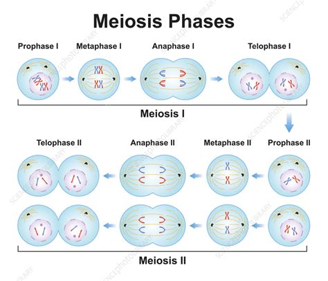
Meiosis, the specialized type of cell division that produces gametes (sperm and egg cells), is a complex process involving two successive divisions: Meiosis I and Meiosis II. Understanding the distinct stages of each division is crucial for grasping the fundamental principles of genetics and inheritance. This guide provides a comprehensive overview of how to identify the stages of meiosis on a diagram, focusing on the key morphological changes in chromosomes and the cell. We'll explore the characteristics of each stage, emphasizing the visual cues that distinguish them.
Meiosis I: Reductional Division
Meiosis I is the reductional division, reducing the chromosome number from diploid (2n) to haploid (n). This ensures that when gametes fuse during fertilization, the resulting zygote maintains the correct diploid chromosome number. Let's dissect the stages:
1. Prophase I: A Detailed Look
Prophase I is the longest and most complex phase of meiosis I. It's characterized by several crucial events:
-
Chromatin Condensation: The replicated chromosomes, each consisting of two sister chromatids joined at the centromere, begin to condense and become visible under a microscope. This is a key visual indicator of the onset of prophase I. Look for long, thread-like structures gradually thickening.
-
Synapsis and Formation of Bivalents: Homologous chromosomes (one from each parent) pair up, a process called synapsis. These paired homologous chromosomes are called bivalents or tetrads (because they contain four chromatids). This is a defining characteristic of Prophase I and crucial for identifying this stage. The pairing is exceptionally tight, creating a complex structure.
-
Crossing Over (Recombination): Non-sister chromatids within the bivalent exchange segments of DNA. This is a vital process that generates genetic diversity by shuffling alleles between homologous chromosomes. On a diagram, crossing over is often depicted as an "X"-shaped structure called a chiasma where the chromatids have exchanged material. Identifying chiasmata is a strong indicator of late Prophase I.
-
Nuclear Envelope Breakdown: Towards the end of Prophase I, the nuclear envelope begins to fragment, allowing the chromosomes to move freely within the cell. The nucleolus also typically disappears.
2. Metaphase I: Alignment on the Metaphase Plate
In Metaphase I, the bivalents align along the metaphase plate, an imaginary plane equidistant from the two poles of the cell. The key visual characteristic is the arrangement of bivalents, not individual chromosomes as in mitosis. The orientation of each bivalent is random, contributing to independent assortment, another source of genetic variation. Look for a line of paired chromosomes positioned across the cell's center.
3. Anaphase I: Separation of Homologous Chromosomes
Anaphase I marks the separation of homologous chromosomes. This is a crucial distinguishing feature from Anaphase II. Sister chromatids remain attached at the centromere. Instead, it's the entire homologous chromosomes that move to opposite poles of the cell, pulled by spindle fibers. Observe the movement of each paired chromosome toward opposite ends.
4. Telophase I and Cytokinesis: Two Haploid Daughter Cells
Telophase I involves the arrival of chromosomes at the poles. The chromosomes may decondense slightly, and a nuclear envelope may reform around each set. Cytokinesis, the division of the cytoplasm, follows Telophase I, resulting in two haploid daughter cells. Each daughter cell contains only one chromosome from each homologous pair, reducing the chromosome number by half. The key observation here is the formation of two separate cells, each with a haploid chromosome number.
Meiosis II: Equational Division
Meiosis II is similar to mitosis but starts with haploid cells. It separates sister chromatids, producing four haploid daughter cells.
1. Prophase II: Chromosomes Condense Again
Prophase II begins with the condensation of chromosomes. Since the homologous chromosomes have already separated in Meiosis I, Prophase II lacks synapsis and crossing over. This is a key distinction from Prophase I. Look for the chromosomes condensing, similar to what is seen in mitotic Prophase.
2. Metaphase II: Individual Chromosomes Align
In Metaphase II, individual chromosomes, each composed of two sister chromatids, align at the metaphase plate. This is the significant difference between Metaphase I and II. In Metaphase I, it's the bivalents (paired homologous chromosomes) that align; in Metaphase II, it's individual chromosomes.
3. Anaphase II: Separation of Sister Chromatids
Anaphase II is characterized by the separation of sister chromatids. This is the crucial event that differentiates it from Anaphase I. The sister chromatids are pulled apart by spindle fibers toward opposite poles. Each chromatid is now considered a separate chromosome.
4. Telophase II and Cytokinesis: Four Haploid Daughter Cells
In Telophase II, chromosomes arrive at the poles. The nuclear envelope reforms, and the chromosomes decondense. Cytokinesis follows, resulting in four haploid daughter cells. These cells are genetically distinct from each other due to crossing over and independent assortment during Meiosis I. The final observation is the generation of four distinct cells, each with half the original chromosome number.
Key Differences: A Summary Table
| Stage | Meiosis I | Meiosis II |
|---|---|---|
| Prophase | Synapsis, crossing over, bivalents formed | Chromosome condensation |
| Metaphase | Bivalents align at metaphase plate | Individual chromosomes align |
| Anaphase | Homologous chromosomes separate | Sister chromatids separate |
| Telophase | Two haploid daughter cells formed | Four haploid daughter cells formed |
| Chromosome # | Reduced from diploid to haploid | Remains haploid |
Practical Tips for Identifying Meiosis Stages on a Diagram
-
Look for the number of chromosomes: Meiosis I starts with a diploid number and ends with a haploid number. Meiosis II starts and ends with a haploid number.
-
Examine the arrangement of chromosomes: In Metaphase I, paired chromosomes are aligned; in Metaphase II, individual chromosomes are aligned.
-
Check for the presence of chiasmata: Chiasmata are indicative of crossing over in Prophase I.
-
Observe the separation of chromosomes: In Anaphase I, homologous chromosomes separate; in Anaphase II, sister chromatids separate.
-
Count the number of cells: Meiosis I produces two cells, while Meiosis II produces four.
Conclusion
Successfully identifying the stages of meiosis on a diagram requires a thorough understanding of the key events in each phase. By carefully observing the chromosome arrangement, the number of chromosomes, and the separation of homologous chromosomes and sister chromatids, you can accurately distinguish between the various stages of both Meiosis I and Meiosis II. Remember, the careful study of diagrams and a solid grasp of the fundamental concepts of meiosis will make this identification process efficient and accurate. This detailed breakdown provides a robust foundation for understanding this intricate and vital process. Remember to practice with various diagrams to solidify your understanding and build confidence in your analysis.
Latest Posts
Latest Posts
-
What Is The Bitrate Of A System
Apr 03, 2025
-
Label The Structures Of The Ankle And Foot
Apr 03, 2025
-
Cpr Practice Test 25 Questions And Answers
Apr 03, 2025
-
You Are Driving Too Slowly If You
Apr 03, 2025
-
Potential Buyers Within A Market Segment Should Be
Apr 03, 2025
Related Post
Thank you for visiting our website which covers about Identify The Stages Of Meiosis On The Diagram . We hope the information provided has been useful to you. Feel free to contact us if you have any questions or need further assistance. See you next time and don't miss to bookmark.
