Label The Bones Of The Skull In Midsagittal View
Breaking News Today
Mar 28, 2025 · 6 min read
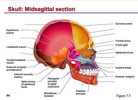
Table of Contents
- Label The Bones Of The Skull In Midsagittal View
- Table of Contents
- Labeling the Bones of the Skull in Midsagittal View: A Comprehensive Guide
- The Midsagittal Plane: A Key Perspective
- Cranial Bones in Midsagittal View
- 1. Frontal Bone: The Forehead's Foundation
- 2. Parietal Bones: The Cranial Dome's Major Players
- 3. Occipital Bone: The Posterior Foundation
- 4. Sphenoid Bone: The Keystone of the Skull Base
- 5. Ethmoid Bone: A Key Component of the Nasal Cavity
- Facial Bones in Midsagittal View
- 1. Vomer: The Nasal Septum's Foundation
- 2. Mandible: The Lower Jaw
- 3. Maxilla: The Upper Jaw (Partial View)
- Significance of Understanding the Midsagittal Skull View
- Conclusion: A Foundation for Further Exploration
- Latest Posts
- Latest Posts
- Related Post
Labeling the Bones of the Skull in Midsagittal View: A Comprehensive Guide
The human skull, a complex structure composed of 22 bones, provides crucial protection for the brain and houses the sensory organs. Understanding its intricate anatomy is essential for students of anatomy, medicine, and related fields. This comprehensive guide focuses on identifying the bones of the skull as viewed in a midsagittal section, providing detailed descriptions and clarifying potential points of confusion. We'll explore each bone, its location, and its significant features, ensuring a thorough understanding of this vital anatomical region.
The Midsagittal Plane: A Key Perspective
Before delving into the individual bones, it’s crucial to understand the perspective: the midsagittal view. This plane divides the skull into equal left and right halves, revealing the structures along the midline. This view allows us to observe the paired and unpaired bones and their relationships with each other. It's a crucial starting point for understanding the overall architecture of the skull.
Cranial Bones in Midsagittal View
The cranial bones protect the brain. In the midsagittal view, we clearly see several of these bones, showcasing their unique shapes and articulations.
1. Frontal Bone: The Forehead's Foundation
The frontal bone forms the anterior part of the skull, contributing significantly to the forehead and superior orbital rim. In the midsagittal view, we observe its vertical portion, extending from the supraorbital margin (above the eyes) to the coronal suture, where it articulates with the parietal bones. The frontal bone's midsagittal aspect showcases its prominent contribution to the anterior cranial fossa, a crucial part of the brain's protective cavity. Look for the frontal crest, a ridge providing attachment for the falx cerebri, a dural fold separating the cerebral hemispheres. The foramen cecum, a small opening at the base of the frontal crest, is also visible in this view, although its significance is primarily related to embryological development.
2. Parietal Bones: The Cranial Dome's Major Players
The parietal bones, two in number, form the majority of the superior and lateral portions of the cranium. In the midsagittal view, only a narrow strip of each parietal bone is visible. The sagittal suture, a significant fibrous joint, is clearly visible separating the two parietal bones. Note the smooth surface of the parietal bones, reflecting their role in protecting the underlying brain tissue. Observe how they articulate with the frontal bone anteriorly (via the coronal suture) and with the occipital bone posteriorly (via the lambdoid suture). The superior and inferior temporal lines, providing attachment points for muscles, are less prominent in this view.
3. Occipital Bone: The Posterior Foundation
The occipital bone forms the posterior and inferior portions of the cranium. The midsagittal view provides a clear view of the external occipital protuberance, a prominent bony landmark easily palpable in most individuals. The internal occipital crest is visible internally, indicating the location of the falx cerebri attachment. The foramen magnum, a large opening at the base of the occipital bone, is prominently displayed in this view. This critical foramen allows passage of the spinal cord from the brain. Note the occipital condyles, which articulate with the first cervical vertebra (atlas), supporting the head's weight. The basilar part of the occipital bone, extending anteriorly towards the sphenoid bone, can also be identified.
4. Sphenoid Bone: The Keystone of the Skull Base
The sphenoid bone, a complex, butterfly-shaped bone, contributes significantly to the skull base. In the midsagittal view, its central component, the body of the sphenoid, is clearly visible. The sella turcica, a saddle-shaped depression housing the pituitary gland, is a striking feature seen in this view. The dorsum sellae, the posterior wall of the sella turcica, is also easily identifiable. The clivus, a sloping portion extending downwards from the dorsum sellae towards the foramen magnum, is another prominent characteristic. Although parts of the greater and lesser wings are visible in the midsagittal section, their full structure is better appreciated in other views.
5. Ethmoid Bone: A Key Component of the Nasal Cavity
The ethmoid bone, a delicate bone situated within the nasal cavity, is partially visible in the midsagittal view. The crista galli, a bony projection extending superiorly, serves as an attachment point for the falx cerebri. The cribriform plate, perforated with numerous foramina (holes) for olfactory nerves, is also seen, but its full structure is best appreciated from other perspectives.
Facial Bones in Midsagittal View
The facial bones provide structural support and shape to the face. In midsagittal view, several key facial bones are visible.
1. Vomer: The Nasal Septum's Foundation
The vomer, a thin, ploughshare-shaped bone, forms the posterior and inferior part of the nasal septum. Its midsagittal view shows its complete structure, highlighting its crucial role in separating the nasal passages.
2. Mandible: The Lower Jaw
The mandible, the only movable bone in the skull, is visible in a midsagittal section. The mandibular symphysis, where the two halves of the mandible fuse during development, is centrally located and often shows a faint ridge in adults. The alveolar process, containing the sockets for the lower teeth, can also be observed. The mental foramen, a small opening providing passage for nerves and blood vessels, is less prominent but may be visible depending on the section.
3. Maxilla: The Upper Jaw (Partial View)
While the full extent of the maxilla is not visible in the midsagittal view, portions of it may appear, showcasing its contribution to the hard palate and the alveolar process for upper teeth.
Significance of Understanding the Midsagittal Skull View
A thorough understanding of the skull's bones in midsagittal view is vital for several reasons:
- Clinical Applications: Radiological images, particularly midline sagittal views, are frequently utilized in diagnosing skull fractures, brain tumors, and other pathologies. Accurate bone identification is crucial for interpretation.
- Surgical Planning: Neurosurgeons and other specialists require detailed knowledge of skull anatomy for planning and executing complex surgeries.
- Forensic Anthropology: Skull fragments found in forensic investigations often require identification of individual bones to ascertain the identity and circumstances of death.
- Anatomical Education: The midsagittal view provides a fundamental framework for understanding the more complex three-dimensional arrangement of the skull bones.
Conclusion: A Foundation for Further Exploration
This guide provides a comprehensive overview of the skull bones visible in a midsagittal view. While this perspective offers crucial insights, exploring additional views (e.g., lateral, superior, inferior) is essential for a complete understanding of the skull’s intricate anatomy. This comprehensive analysis provides a strong foundation for further exploration and learning, empowering you to confidently identify and understand the complex structure of the human skull. Remember that consistent study and visualization, ideally with physical models or anatomical charts, are crucial to mastering this challenging yet rewarding aspect of human anatomy.
Latest Posts
Latest Posts
-
Antibodies Are Produced From Which Cells Quizlet
Mar 31, 2025
-
Ethics And Law In Leadership Edapt Quizlet
Mar 31, 2025
-
Compare And Contrast Indentured Servanthood And Slavery
Mar 31, 2025
-
Ac Theory Level 2 Lesson 8 Quizlet
Mar 31, 2025
-
Dts Personal Leave With Official Travel Quizlet
Mar 31, 2025
Related Post
Thank you for visiting our website which covers about Label The Bones Of The Skull In Midsagittal View . We hope the information provided has been useful to you. Feel free to contact us if you have any questions or need further assistance. See you next time and don't miss to bookmark.
