Label The Cranial Dura Septa In The Figure.
Breaking News Today
Mar 26, 2025 · 6 min read
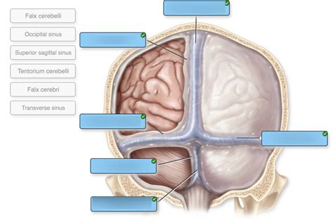
Table of Contents
Label the Cranial Dura Septa in the Figure: A Comprehensive Guide
The dura mater, the outermost layer of the meninges, is not a simple, continuous sheet. Instead, it forms several important septa, or partitions, that extend inward to compartmentalize the cranial cavity and support the brain. Understanding the location and function of these dural septa is crucial for anyone studying neuroanatomy. This article will provide a detailed explanation of each septum, guiding you through their identification and significance. We will focus on accurately labeling these structures and exploring their clinical relevance.
The Major Dural Septa: Falx Cerebri, Tentorium Cerebelli, Falx Cerebelli, and Diaphragma Sellae
The cranial dura mater is composed of two layers: the periosteal layer (outer) and the meningeal layer (inner). The septa are formed by inward projections of the meningeal layer. Let's explore each major septum:
1. Falx Cerebri: Dividing the Cerebral Hemispheres
The falx cerebri is arguably the most prominent dural septum. Its sickle-shaped structure extends vertically from the crista galli of the ethmoid bone (anteriorly) to the internal occipital protuberance (posteriorly). This large, crescent-shaped fold sits within the longitudinal fissure, separating the two cerebral hemispheres.
Key Features & Labeling:
- Superior margin: Attached to the inner surface of the skull along the sagittal suture.
- Inferior margin: Free edge that arches over the corpus callosum.
- Anterior attachment: Crista galli of the ethmoid bone.
- Posterior attachment: Internal occipital protuberance.
- Superior sagittal sinus: Located within the superior margin, a major venous channel draining blood from the brain.
- Inferior sagittal sinus: Smaller sinus located along the inferior margin.
Clinical Significance: The falx cerebri's location makes it susceptible to injury in traumatic brain injury (TBI). Hematoma formation within the falx cerebri can lead to significant neurological deficits, requiring immediate medical intervention.
2. Tentorium Cerebelli: Separating Cerebrum from Cerebellum
The tentorium cerebelli is a large, tent-like structure that separates the occipital lobes of the cerebrum from the cerebellum. Its shape is crucial for protecting the cerebellum and brainstem from the weight and movement of the cerebrum.
Key Features & Labeling:
- Superior surface: Concave and accommodates the occipital lobes.
- Inferior surface: Concave and rests upon the cerebellum.
- Anterior attachment: Clinoid processes of the sphenoid bone.
- Posterior attachment: Transverse sinus and internal occipital crest.
- Free posterior border: Forms the incisura tentorii, a gap through which the brainstem passes.
- Transverse sinuses: Located along the attachment to the occipital bone.
Clinical Significance: The tentorium cerebelli's rigid structure can cause herniation of the brainstem in cases of increased intracranial pressure (ICP). This life-threatening condition, known as uncal herniation (specifically involving the uncus of the temporal lobe), can result in coma and death. The incisura tentorii is a key anatomical landmark in understanding this process.
3. Falx Cerebelli: Separating the Cerebellar Hemispheres
Much smaller than the falx cerebri, the falx cerebelli is a small, sickle-shaped fold of dura that separates the two cerebellar hemispheres. It extends inferiorly from the internal occipital crest and attaches to the posterior aspect of the foramen magnum.
Key Features & Labeling:
- Attachment: Internal occipital crest.
- Inferior end: Attaches to the posterior aspect of the foramen magnum.
- Occipital sinus: Lies within its posterior border.
Clinical Significance: While less frequently involved in clinical scenarios compared to the falx cerebri and tentorium cerebelli, the falx cerebelli still plays a role in compartmentalizing the cerebellum and protecting against potential injury.
4. Diaphragma Sellae: Enclosing the Pituitary Gland
The diaphragma sellae is a small, circular dural fold that forms a roof over the sella turcica of the sphenoid bone. This structure encloses the pituitary gland, protecting it from surrounding structures.
Key Features & Labeling:
- Attachment: Edges of the sella turcica.
- Central opening: Allows for the passage of the infundibulum (pituitary stalk) which connects the pituitary gland to the hypothalamus.
Clinical Significance: The diaphragma sellae is important in pituitary adenoma management. Understanding its anatomical relationship to the pituitary gland is crucial for neurosurgical procedures.
Less Prominent Dural Reflections and their Clinical Importance
While the four septa described above are the most prominent, several smaller dural reflections contribute to the complex structure of the cranial dura. These less prominent structures, although smaller, still play important roles in the brain's support and protection, and can be relevant in certain clinical situations. Here are some key ones to consider:
-
Cavernous sinus: While not a septum per se, it's an important dural venous plexus located on either side of the sella turcica. It houses significant cranial nerves (III, IV, V1, V2, VI) and the internal carotid artery. Infections (e.g., cavernous sinus thrombosis) or aneurysms in this region can have devastating consequences.
-
Superior and inferior petrosal sinuses: These sinuses are located within the dural folds near the petrous portion of the temporal bone and contribute to the drainage of venous blood from the brain. Their close proximity to important cranial nerves warrants attention in clinical settings.
-
Straight sinus: Formed by the confluence of the inferior sagittal sinus and the great cerebral vein of Galen. It is part of the major venous drainage pathway of the brain.
-
Confluence of sinuses: The junction point of the superior sagittal sinus, transverse sinuses, and straight sinus. This area is a crucial confluence of venous drainage.
Labeling a Figure: Practical Tips
When labeling a figure of the cranial dura septa, accuracy is paramount. Use clear and concise labels, avoiding ambiguity. Ensure your labels are aligned with the anatomical structures they represent. Consider using different colors or line styles to distinguish between different septa for enhanced clarity. High-quality anatomical illustrations are essential. It is helpful to have a side-by-side comparison with high quality anatomical images for proper identification and labelling.
Here's a suggested approach:
- Obtain a high-resolution anatomical image: A clear image of the cranial dura and its septa is crucial.
- Identify key landmarks: Begin by identifying easily recognizable landmarks like the crista galli, sella turcica, and internal occipital protuberance.
- Systematic approach: Label the major septa (falx cerebri, tentorium cerebelli, falx cerebelli, and diaphragma sellae) first.
- Add minor structures: Once the major septa are labeled, add the lesser structures such as the sinuses.
- Careful annotation: Use precise labeling, avoiding ambiguity. Use a legend or key to explain any abbreviations or symbols used.
Clinical Relevance and Deeper Dive
The dural septa are not merely anatomical curiosities. Their clinical relevance is significant. Understanding their roles in protecting the brain and their susceptibility to injury is crucial for diagnosing and managing various neurological conditions. Further exploration into specific conditions linked to these septa is warranted:
-
Subdural hematoma: Bleeding between the dura mater and the arachnoid mater. Often caused by trauma, it can compress the brain and cause serious neurological consequences.
-
Epidural hematoma: Bleeding between the skull and the dura mater. Typically caused by a skull fracture, it can rapidly expand and compress the brain, requiring immediate surgical intervention.
-
Meningitis: Inflammation of the meninges, which can involve the dura mater. It can be caused by bacterial, viral, or fungal infections.
-
Brain herniation: The protrusion of brain tissue through an opening in the dura mater. This is a life-threatening condition typically associated with increased ICP.
-
Venous sinus thrombosis: A blood clot in a dural venous sinus. It can be caused by various factors, including infection, dehydration, or malignancy.
By understanding the intricate anatomy of the cranial dura septa and their clinical implications, medical professionals can effectively diagnose and manage a wide range of neurological conditions, ensuring optimal patient care. Accurate labeling and identification of these structures during anatomical study are thus essential. The more you practice, the more proficient you will become at recognizing and identifying these crucial structures.
Latest Posts
Latest Posts
-
Obtained By Having Had A Contagious Disease
Mar 29, 2025
-
Choose The Statement Below That Explains What Closing Means
Mar 29, 2025
-
Music With No Literary Basis Is Referred To As
Mar 29, 2025
-
Whats One Main Difference Between Windows And Linux Processes
Mar 29, 2025
-
Label The Structures Of The Lower Respiratory Tract
Mar 29, 2025
Related Post
Thank you for visiting our website which covers about Label The Cranial Dura Septa In The Figure. . We hope the information provided has been useful to you. Feel free to contact us if you have any questions or need further assistance. See you next time and don't miss to bookmark.
