Label The Structures Of The Lower Respiratory Tract.
Breaking News Today
Mar 29, 2025 · 6 min read
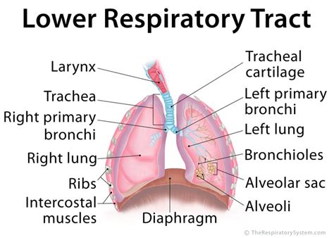
Table of Contents
Label the Structures of the Lower Respiratory Tract: A Comprehensive Guide
The lower respiratory tract, responsible for gas exchange, is a complex system of branching tubes and delicate air sacs. Understanding its intricate anatomy is crucial for comprehending respiratory function and various respiratory diseases. This comprehensive guide provides a detailed walkthrough of the lower respiratory tract's structures, accompanied by clear explanations and helpful labeling to aid learning and comprehension.
The Trachea: The Windpipe's Role
The trachea, or windpipe, is the first major structure of the lower respiratory tract. It's a cartilaginous tube approximately 10-12 cm long and 2 cm in diameter, extending from the larynx (voice box) to the carina, where it bifurcates (splits) into the two main bronchi. Its rigid structure, thanks to C-shaped hyaline cartilage rings, prevents collapse during inhalation and exhalation. The gaps in these rings are bridged by the trachealis muscle and connective tissue, allowing for some flexibility and expansion during swallowing. The inner lining of the trachea, the mucosa, is lined with pseudostratified columnar epithelium containing goblet cells that secrete mucus. This mucus traps inhaled particles, preventing them from reaching the delicate alveoli. Cilia, hair-like projections on the epithelial cells, beat rhythmically to move the mucus upward, towards the pharynx, where it is swallowed or expelled.
Key Features of the Trachea:
- C-shaped hyaline cartilage rings: Provide structural support and prevent collapse.
- Trachealis muscle: Allows for expansion and flexibility during swallowing.
- Mucosa: Contains goblet cells (mucus secretion) and cilia (mucus transport).
- Carina: The point of bifurcation into the main bronchi.
The Bronchial Tree: Branching Out to the Alveoli
The trachea branches into two main (primary) bronchi, one for each lung. These bronchi then further subdivide into progressively smaller branches, forming the bronchial tree. This branching pattern resembles an inverted tree, with the trachea as the trunk and the alveoli as the leaves. As the branches get smaller, the amount of cartilage decreases, and the proportion of smooth muscle increases. This smooth muscle plays a crucial role in regulating airflow by bronchodilation (widening) and bronchoconstriction (narrowing).
Bronchial Tree Subdivision:
- Main (primary) bronchi: Two large branches, one for each lung.
- Lobar (secondary) bronchi: Three on the right lung, two on the left, supplying each lobe.
- Segmental (tertiary) bronchi: Supply bronchopulmonary segments within each lobe.
- Bronchioles: Smaller branches lacking cartilage, with increased smooth muscle.
- Terminal bronchioles: The smallest conducting bronchioles.
- Respiratory bronchioles: The transition zone where gas exchange begins.
- Alveolar ducts: Lead to alveolar sacs.
- Alveolar sacs: Clusters of alveoli.
- Alveoli: Tiny air sacs where gas exchange occurs.
The Lungs: The Site of Gas Exchange
The lungs are the primary organs of the respiratory system, responsible for the vital process of gas exchange – the uptake of oxygen (O2) and the release of carbon dioxide (CO2). Each lung is enclosed in a double-layered pleura, a serous membrane that produces pleural fluid, acting as a lubricant to reduce friction during breathing. The outer layer, the parietal pleura, lines the thoracic cavity, while the inner layer, the visceral pleura, adheres directly to the lung surface.
Lung Structure: Lobes and Segments
- Right Lung: Larger and has three lobes (superior, middle, inferior).
- Left Lung: Smaller and has two lobes (superior, inferior), due to the heart's location.
- Bronchopulmonary Segments: Each lobe is further subdivided into bronchopulmonary segments, functionally independent units supplied by a segmental bronchus and its associated pulmonary artery and vein. These segments are clinically important because they can be surgically removed without affecting the rest of the lung.
The Alveoli: The Tiny Air Sacs of Gas Exchange
The alveoli are the functional units of the lungs. These tiny, thin-walled air sacs are clustered together like grapes, forming alveolar sacs. Their thin walls, composed primarily of a single layer of type I alveolar cells (squamous epithelial cells), are ideal for efficient gas exchange. Type II alveolar cells secrete pulmonary surfactant, a lipoprotein that reduces surface tension in the alveoli, preventing their collapse during exhalation. Alveoli are surrounded by a network of capillaries, allowing for close proximity between air and blood, facilitating efficient diffusion of gases.
The Alveolar-Capillary Membrane: The Site of Gas Exchange
The alveolar-capillary membrane (respiratory membrane) is the barrier between the alveolar air and the blood in the pulmonary capillaries. It consists of:
- Alveolar epithelium (type I cells): Single layer of squamous cells.
- Alveolar basement membrane: Thin layer of connective tissue.
- Capillary basement membrane: May fuse with the alveolar basement membrane.
- Capillary endothelium: Single layer of endothelial cells.
This extremely thin membrane allows for rapid diffusion of oxygen from the alveoli into the blood and carbon dioxide from the blood into the alveoli.
Pulmonary Circulation: Blood Supply to the Lungs
The lungs receive a dual blood supply:
- Pulmonary Circulation: Low-pressure, high-volume circulation carrying deoxygenated blood from the right ventricle of the heart to the pulmonary capillaries for oxygenation, and then returning oxygenated blood to the left atrium.
- Bronchial Circulation: High-pressure, low-volume circulation originating from the aorta, supplying oxygenated blood to the lung tissue itself (bronchi, bronchioles, connective tissue).
Clinical Significance and Respiratory Diseases
Understanding the structure of the lower respiratory tract is essential for diagnosing and treating various respiratory diseases. Conditions affecting different parts of the respiratory system manifest with unique symptoms and require specific treatment strategies. For instance:
- Asthma: Affects the bronchioles, causing inflammation and bronchoconstriction.
- Pneumonia: Infection of the alveoli, leading to impaired gas exchange.
- Emphysema: Destruction of alveolar walls, reducing the surface area for gas exchange.
- Lung Cancer: Can originate in any part of the lower respiratory tract, including bronchi and alveoli.
- Pulmonary Embolism: Blockage of pulmonary arteries, often by a blood clot.
- Pleurisy (Pleuritis): Inflammation of the pleura, causing chest pain and difficulty breathing.
Accurate labeling of these structures is crucial for medical professionals, providing a clear understanding of the location and extent of these pathologies.
Conclusion: Mastering the Lower Respiratory Tract's Anatomy
This detailed guide provides a thorough overview of the lower respiratory tract's structures, from the trachea to the alveoli. The intricate branching pattern of the bronchial tree, the unique structure of the alveoli, and the vital role of the pulmonary circulation in gas exchange highlight the complexity and importance of this system. A strong understanding of this anatomy is crucial for both comprehending respiratory physiology and diagnosing and treating related diseases. Continued study and referencing detailed anatomical diagrams are highly recommended for a comprehensive mastery of this topic. Remember to consult reliable medical textbooks and resources for further in-depth learning and clarification.
Latest Posts
Latest Posts
-
Match The Term With Its Correct Description
Apr 01, 2025
-
Rn Medical Surgical Online Practice 2023 A
Apr 01, 2025
-
Yo Querer Ver Una Pelicula Horror
Apr 01, 2025
-
What Are The Qualities Of A Good Hypothesis
Apr 01, 2025
-
What Tool Detangles And Styles Wigs Hairpieces And Hair Additions
Apr 01, 2025
Related Post
Thank you for visiting our website which covers about Label The Structures Of The Lower Respiratory Tract. . We hope the information provided has been useful to you. Feel free to contact us if you have any questions or need further assistance. See you next time and don't miss to bookmark.
