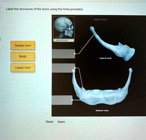
Label The Structures Of The Bone Using The Hints Provided
Breaking News Today
Apr 02, 2025 · 7 min read

Table of Contents
Label the Structures of the Bone: A Comprehensive Guide
Understanding bone structure is fundamental to comprehending the skeletal system's function and overall human anatomy. This detailed guide will walk you through labeling the various structures of a bone, using hints to aid in identification. We'll explore both macroscopic and microscopic features, providing a comprehensive understanding for students, enthusiasts, and anyone curious about the amazing complexity of bones.
Macroscopic Bone Structures: The Big Picture
Before diving into microscopic details, let's familiarize ourselves with the major components visible to the naked eye. A typical long bone, like the femur or humerus, serves as an excellent example to illustrate these structures.
1. Diaphysis (Shaft): The Long Part of the Bone
The diaphysis is the long, cylindrical shaft of the bone. It's primarily composed of compact bone, providing strength and structural support. Think of it as the main, load-bearing part of the bone.
Hint: Look for the longest, straightest part of the bone. That's your diaphysis!
2. Epiphysis (Ends): The Growth Plates and Articulating Surfaces
The epiphysis refers to the two ends of a long bone. These expanded portions are crucial for articulation—the joining of bones at a joint. They contain spongy bone, which is lighter yet strong enough to withstand stress. Importantly, the epiphysis houses the epiphyseal plate (growth plate) during childhood and adolescence.
Hint: Look for the wider, often rounded, sections at the ends of the bone where it connects to other bones. These are the epiphyses.
3. Metaphysis: The Transition Zone
The metaphysis is the region where the diaphysis meets the epiphysis. In growing bones, this area contains the epiphyseal plate (growth plate), a cartilaginous region responsible for longitudinal bone growth. Once growth ceases, the epiphyseal plate ossifies, forming the epiphyseal line.
Hint: Look for the slightly flared area between the diaphysis and epiphysis. This area often shows a distinct line (epiphyseal line) in adult bones.
4. Articular Cartilage: Cushioning the Joints
Covering the epiphyses where bones meet is the articular cartilage. This smooth, hyaline cartilage reduces friction and acts as a shock absorber during joint movement. It's essential for maintaining joint health and preventing wear and tear.
Hint: This is a very smooth, glistening surface at the ends of the bones where they articulate.
5. Periosteum: The Tough Outer Membrane
The periosteum is a tough, fibrous connective tissue membrane that covers the outer surface of the bone (except for the articular cartilage). It contains blood vessels, nerves, and osteoblasts (bone-forming cells), playing a crucial role in bone growth, repair, and nutrition.
Hint: Imagine it as the bone's protective outer "skin." It's a tough, whitish layer.
6. Endosteum: Lining the Inner Cavity
The endosteum is a thin membrane that lines the inner surface of the medullary cavity (the hollow space within the diaphysis). It contains osteoblasts and osteoclasts (bone-resorbing cells), involved in bone remodeling.
Hint: It's the inner lining of the hollow shaft of the bone.
7. Medullary Cavity: The Marrow-Filled Space
The medullary cavity is the hollow space within the diaphysis. In adults, this cavity primarily contains yellow bone marrow, composed mostly of fat cells. In children, it contains red bone marrow, which is involved in blood cell formation (hematopoiesis).
Hint: This is the hollow space inside the diaphysis.
8. Nutrient Foramen: Entry Point for Blood Vessels
The nutrient foramen is a small opening in the diaphysis that allows blood vessels to enter the medullary cavity, supplying nutrients to the bone tissue.
Hint: Look for small holes along the shaft of the bone.
9. Processes, Tuberosities, and Condyles: Attachment Points for Muscles and Ligaments
Various bony projections, such as processes, tuberosities, and condyles, serve as attachment points for muscles, tendons, and ligaments. Their shapes and locations are highly variable depending on the specific bone.
Hint: These are the bumpy, rough areas on the bone's surface where muscles and ligaments attach.
Microscopic Bone Structures: Zooming In
Now let's explore the microscopic organization of bone tissue. This level of detail reveals the intricate arrangement of cells and extracellular matrix that gives bones their strength and resilience.
10. Osteocytes: The Bone Cells
Osteocytes are mature bone cells residing within lacunae (small spaces) within the bone matrix. They maintain the bone tissue and are responsible for sensing mechanical stress on the bone.
Hint: These are the main "living" cells within the bone structure.
11. Lacunae: The Osteocyte Homes
Lacunae are small spaces within the bone matrix where osteocytes reside. They're interconnected by canaliculi, tiny channels that allow communication and nutrient exchange between osteocytes.
Hint: These are the small spaces containing the osteocytes.
12. Canaliculi: Connecting the Osteocytes
Canaliculi are microscopic canals that connect lacunae, allowing for the passage of nutrients, waste products, and communication signals between osteocytes.
Hint: These are the tiny channels radiating from the lacunae.
13. Lamellae: Concentric Rings of Bone Matrix
Lamellae are concentric rings of bone matrix that surround the central canal (Haversian canal) in compact bone. These rings give compact bone its characteristic organized structure.
Hint: Look for the circular rings of bone material.
14. Haversian Canal (Central Canal): The Blood Vessel Highway
The Haversian canal (also known as the central canal) runs longitudinally through the compact bone, containing blood vessels and nerves that supply the osteocytes.
Hint: This is the central channel running through the lamellae.
15. Volkmann's Canals: Connecting the Haversian Canals
Volkmann's canals are transverse canals that connect adjacent Haversian canals, providing additional pathways for blood vessels and nerves.
Hint: These are the canals that connect the central canals to each other and to the periosteum.
16. Spongy Bone (Cancellous Bone): A Network of Trabeculae
Spongy bone, also known as cancellous bone, is a porous bone tissue found in the epiphyses of long bones and the interior of other bones. It's composed of a network of trabeculae (thin, bony struts), creating a lightweight yet strong structure. Red bone marrow is often found within the spaces of spongy bone.
Hint: This is the more porous type of bone tissue, often described as having a honeycombed appearance.
17. Trabeculae: The Supporting Struts of Spongy Bone
Trabeculae are the thin, bony struts that make up spongy bone. Their arrangement is influenced by the stresses placed upon the bone, providing strength and support where it's needed most.
Hint: These are the small beams or struts forming the framework of spongy bone.
18. Bone Matrix: The Extracellular Substance
The bone matrix is the extracellular substance that makes up the majority of bone tissue. It's composed primarily of collagen fibers and mineral salts, providing both flexibility and rigidity to the bone.
Hint: This is the non-cellular component of the bone, giving it its strength and structure.
Putting it All Together: A Practical Approach
The best way to truly grasp bone structure is through hands-on learning. If possible, use anatomical models, diagrams, or even real bone specimens (with proper safety precautions and guidance) to identify the structures described above.
Use the hints provided as a guide, carefully examine the bone, and try to correlate the macroscopic features with their microscopic components. For example, understand how the Haversian canals in compact bone contribute to the overall strength of the diaphysis.
Remember: Bone structure is complex and intricate, but by breaking it down into smaller, manageable components and utilizing the hints given, you can gain a comprehensive understanding of this vital part of the human body. This detailed approach not only allows for successful labeling but also fosters a deeper appreciation for the remarkable engineering of the skeletal system.
Beyond Labeling: Understanding Bone Function and Pathology
Understanding the structures of a bone is crucial, but it's equally important to appreciate their function and how problems can arise. Here's a brief look:
- Bone Remodeling: A continuous process of bone breakdown (resorption by osteoclasts) and bone formation (by osteoblasts) that maintains bone health and adapts to mechanical stress.
- Fractures: Bone breaks, ranging in severity from hairline cracks to complete shattering. Understanding bone structure is essential for diagnosis and treatment.
- Osteoporosis: A condition characterized by reduced bone density, making bones more fragile and prone to fracture. Knowledge of bone structure helps explain the increased risk of fracture in osteoporosis.
- Osteoarthritis: Degenerative joint disease affecting articular cartilage, leading to pain, stiffness, and limited movement. The interplay between bone and cartilage is crucial to understand this condition.
By gaining a thorough understanding of the structures of bone, we can gain insight into various skeletal conditions and appreciate the complex interplay of cells, tissues, and mechanical forces that maintain skeletal health. This knowledge extends beyond simple labeling and provides a foundation for further exploration of this fascinating field. Through continued learning and exploration, we can develop a much deeper understanding of this vital part of the human body.
Latest Posts
Latest Posts
-
What Is The Bitrate Of A System
Apr 03, 2025
-
Label The Structures Of The Ankle And Foot
Apr 03, 2025
-
Cpr Practice Test 25 Questions And Answers
Apr 03, 2025
-
You Are Driving Too Slowly If You
Apr 03, 2025
-
Potential Buyers Within A Market Segment Should Be
Apr 03, 2025
Related Post
Thank you for visiting our website which covers about Label The Structures Of The Bone Using The Hints Provided . We hope the information provided has been useful to you. Feel free to contact us if you have any questions or need further assistance. See you next time and don't miss to bookmark.
