Match Each Picture With The Correct Stage Of Mitosis.
Breaking News Today
Mar 20, 2025 · 6 min read
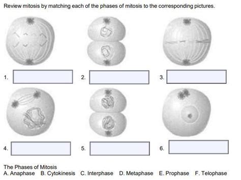
Table of Contents
Matching Pictures with the Correct Stage of Mitosis: A Comprehensive Guide
Mitosis, the process of cell division resulting in two identical daughter cells, is a fundamental concept in biology. Understanding its stages – prophase, prometaphase, metaphase, anaphase, and telophase – is crucial for grasping the mechanics of life itself. This detailed guide provides a comprehensive overview of each mitotic stage, accompanied by visual aids (imagine pictures here, representing each stage, which would be inserted in a real blog post) to aid in your understanding and identification. We'll delve into the key characteristics of each phase, focusing on the observable changes within the cell's structure and the movement of its chromosomes.
Understanding the Fundamentals of Mitosis
Before diving into the specifics of each stage, let's establish a foundational understanding. Mitosis ensures the accurate replication and distribution of genetic material, ensuring that each daughter cell receives a complete and identical set of chromosomes. This is essential for growth, repair, and asexual reproduction in eukaryotic organisms. The process is tightly regulated, preventing errors that could lead to genetic instability and potentially cancerous growth.
The entire process is a continuous one, but for clarity, we divide it into distinct stages: prophase, prometaphase, metaphase, anaphase, and telophase. These stages are characterized by distinct morphological changes visible under a microscope. Remember, the transition between stages is gradual, and the precise boundaries can sometimes be subjective depending on the cell type and the imaging techniques used.
Prophase: The Initial Stage of Chromosomal Condensation
(Imagine a picture of prophase here showing condensed chromosomes)
Prophase marks the beginning of mitosis. Here, several crucial events take place:
-
Chromatin Condensation: The loose, thread-like chromatin fibers, which represent the uncondensed DNA, begin to condense and coil tightly, forming visible chromosomes. Each chromosome consists of two identical sister chromatids joined at the centromere. This condensation is essential for accurate segregation during later stages.
-
Nuclear Envelope Breakdown: The nuclear envelope, the membrane surrounding the nucleus, starts to break down. This allows the chromosomes to access the spindle fibers, crucial for their movement.
-
Spindle Formation: The mitotic spindle, a structure composed of microtubules (protein filaments), begins to form. This structure originates from the centrosomes, which duplicate and migrate to opposite poles of the cell during prophase. The spindle fibers will play a critical role in the segregation of chromosomes.
-
Nucleolus Disappearance: The nucleolus, a structure within the nucleus responsible for ribosome production, disappears during prophase.
Key Characteristics of Prophase:
- Distinct, condensed chromosomes are visible.
- The nuclear envelope is disintegrating.
- The mitotic spindle is forming.
- The nucleolus is no longer visible.
Prometaphase: The Chromosomes Attach to the Spindle
(Imagine a picture of prometaphase here showing chromosomes attaching to spindle fibers)
Prometaphase, a transitional phase, bridges the gap between prophase and metaphase. The key event here is the attachment of the chromosomes to the spindle fibers:
-
Kinetochore Formation: Specialized protein structures called kinetochores assemble at the centromeres of each chromosome. These kinetochores serve as the attachment points for the spindle fibers.
-
Chromosome Movement: The chromosomes begin to move towards the center of the cell, although they are not yet perfectly aligned. This movement is driven by the dynamic interactions between the kinetochores and the spindle fibers.
-
Spindle Fiber Growth: The spindle fibers continue to grow and extend from the centrosomes at opposite poles of the cell, filling the cytoplasm.
Key Characteristics of Prometaphase:
- Chromosomes show more defined condensation.
- Kinetochores are visible on the centromeres.
- Chromosomes are actively moving towards the cell's equator.
- Spindle fibers are attached to the kinetochores.
Metaphase: Chromosomes Align at the Metaphase Plate
(Imagine a picture of metaphase here showing chromosomes aligned at the metaphase plate)
Metaphase represents a critical checkpoint in mitosis. Here, the chromosomes achieve perfect alignment:
-
Chromosome Alignment: The chromosomes align at the cell's equator, forming the metaphase plate. This alignment is crucial to ensure that each daughter cell receives an identical set of chromosomes.
-
Spindle Fiber Tension: The spindle fibers exert equal tension on the sister chromatids, pulling them towards opposite poles. This tension is essential for accurate separation in the next stage.
-
Spindle Checkpoint: A crucial checkpoint mechanism ensures that all chromosomes are correctly attached to the spindle fibers before proceeding to anaphase. This checkpoint prevents errors in chromosome segregation.
Key Characteristics of Metaphase:
- Chromosomes are perfectly aligned at the metaphase plate.
- Sister chromatids are held together by the centromere.
- Spindle fibers are under tension, pulling on the chromosomes.
Anaphase: Sister Chromatids Separate
(Imagine a picture of anaphase here showing sister chromatids separating)
Anaphase is the shortest stage of mitosis, yet it's where the most dramatic events occur:
-
Sister Chromatid Separation: The sister chromatids are suddenly separated at the centromere. This separation is driven by the shortening of the spindle fibers. Each chromatid is now considered an independent chromosome.
-
Chromosome Movement: The separated chromosomes move towards opposite poles of the cell, pulled by the retracting spindle fibers. This movement is precisely coordinated to ensure that each pole receives a complete set of chromosomes.
-
Cell Elongation: The cell begins to elongate as the poles move further apart.
Key Characteristics of Anaphase:
- Sister chromatids are separating.
- Chromosomes are moving towards the poles.
- The cell is elongating.
Telophase: Cytokinesis and the Formation of Two Daughter Cells
(Imagine a picture of telophase here showing two newly formed daughter cells)
Telophase marks the final stage of mitosis, where the process concludes:
-
Chromosome Decondensation: The chromosomes reach the poles and begin to decondense, returning to their extended chromatin form.
-
Nuclear Envelope Reformation: A new nuclear envelope forms around each set of chromosomes at each pole, creating two separate nuclei.
-
Nucleolus Reformation: The nucleoli reappear within each newly formed nucleus.
-
Spindle Fiber Disassembly: The mitotic spindle disassembles, its components recycled within the cell.
-
Cytokinesis: Cytokinesis, the division of the cytoplasm, occurs concurrently with telophase, resulting in the formation of two genetically identical daughter cells. In animal cells, a cleavage furrow forms, pinching the cell in two. In plant cells, a cell plate forms, separating the two daughter cells.
Key Characteristics of Telophase:
- Chromosomes decondense.
- Nuclear envelopes reform.
- Nucleoli reappear.
- Cytokinesis occurs, resulting in two daughter cells.
Putting it All Together: A Comprehensive Summary
Mitosis is a complex but elegant process essential for the growth and maintenance of multicellular organisms. By carefully examining the characteristic features of each phase – prophase, prometaphase, metaphase, anaphase, and telophase – we can better appreciate the precision and regulation involved in this fundamental cellular event. Each phase plays a vital role in ensuring the accurate replication and distribution of genetic material, leading to the creation of two genetically identical daughter cells.
Remember, the pictures (which should accompany this text in a real blog post) are crucial for reinforcing your understanding. Comparing your observations of these images to the descriptions provided will significantly enhance your ability to identify the stages of mitosis accurately. Practice identifying the stages in different cell types and under various microscopic techniques; this will hone your understanding of this fundamental biological process. Further research into the molecular mechanisms driving each stage will provide even deeper insight into the intricate world of cellular division.
Latest Posts
Latest Posts
-
What In Broad Terms Is The Definition Of Deviance
Mar 20, 2025
-
B Boying Is Highly Choreographed Group Of Answer Choices True False
Mar 20, 2025
-
Writing An Argumentative Essay About The Nobel Prize In Literature
Mar 20, 2025
-
When You Delete A Folder What Happens
Mar 20, 2025
-
Which Of Fridays Traits Does Crusoe Find Admirable
Mar 20, 2025
Related Post
Thank you for visiting our website which covers about Match Each Picture With The Correct Stage Of Mitosis. . We hope the information provided has been useful to you. Feel free to contact us if you have any questions or need further assistance. See you next time and don't miss to bookmark.
