Nerve Fibers From The Medial Aspect Of Each Eye
Breaking News Today
Mar 20, 2025 · 6 min read
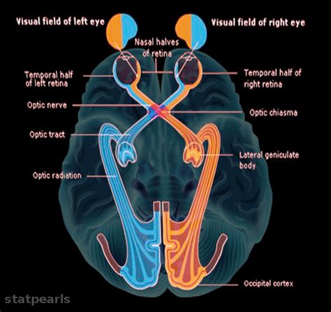
Table of Contents
Nerve Fibers from the Medial Aspect of Each Eye: A Deep Dive into Nasal Retinal Fibers and Their Clinical Significance
The human visual system is a marvel of biological engineering, capable of processing vast amounts of information to create our perception of the world. Understanding the intricate pathways of this system is crucial for diagnosing and treating a wide range of ophthalmologic conditions. This article delves into the fascinating world of nerve fibers originating from the medial aspect of each eye – specifically, the nasal retinal fibers – exploring their anatomy, physiology, and clinical implications.
Anatomy of Nasal Retinal Fibers
The retina, the light-sensitive tissue lining the back of the eye, is composed of millions of photoreceptor cells (rods and cones) that convert light into electrical signals. These signals are then processed by a complex network of retinal neurons before being transmitted to the brain via the optic nerve. The optic nerve, or cranial nerve II, carries these signals from the eye to the optic chiasm, a crucial structure located at the base of the brain.
Nasal retinal fibers, originating from the medial (nasal) half of each retina, hold a unique position in this visual pathway. Unlike the temporal retinal fibers (originating from the lateral half of the retina), these fibers undergo a significant anatomical rearrangement at the optic chiasm. This characteristic is fundamental to our binocular vision.
The Optic Chiasm: A Point of Decussation
At the optic chiasm, approximately half of the nerve fibers from each optic nerve cross over to the opposite side of the brain. It's specifically the nasal retinal fibers that decussate, meaning they cross over to the contralateral side. The temporal retinal fibers, on the other hand, remain ipsilateral, meaning they stay on the same side of the brain. This crossing is essential for creating a unified visual field perception.
This arrangement ensures that information from the left half of the visual field (processed by both eyes' nasal retinas) is relayed to the right side of the brain, and vice-versa. This ensures that the visual cortex, located in the occipital lobe, receives a complete and integrated representation of the visual world.
Pathways Beyond the Optic Chiasm: The Optic Tracts and Lateral Geniculate Nucleus
After the optic chiasm, the nerve fibers continue as the optic tracts. These tracts contain a mixture of ipsilateral temporal fibers and contralateral nasal fibers. The optic tracts terminate primarily in the lateral geniculate nucleus (LGN) of the thalamus, a crucial relay station in the visual pathway. The LGN further processes the visual information before sending it to the visual cortex via the optic radiations.
The Visual Cortex: Processing Visual Information
The visual cortex in the occipital lobe is where the complex process of visual perception takes place. The information from the LGN is meticulously analyzed, allowing us to interpret shapes, colors, movement, and depth perception. The arrangement of nasal and temporal fibers within the optic tracts and the LGN ensures that the visual cortex receives a spatially organized representation of the visual field.
Physiological Significance of Nasal Retinal Fibers
The nasal retinal fibers play a vital role in several aspects of visual function:
-
Binocular Vision: As mentioned earlier, the decussation of nasal fibers is crucial for binocular vision, our ability to see with both eyes simultaneously, creating depth perception and three-dimensional awareness. Without this crossing, our visual perception would be significantly impaired.
-
Peripheral Vision: Nasal retinal fibers contribute significantly to our peripheral vision, allowing us to perceive objects and movements outside our central visual field. This peripheral awareness is essential for navigation, safety, and overall spatial orientation.
-
Visual Field Integration: The precise arrangement and processing of information from nasal and temporal retinal fibers ensure that our brain integrates the visual field seamlessly, producing a unified and coherent visual experience.
Clinical Significance of Nasal Retinal Fibers
Damage to the nasal retinal fibers or their pathways can result in a variety of visual impairments, providing crucial diagnostic information for ophthalmologists and neurologists.
Visual Field Defects: Scotomas and Hemianopsias
Damage anywhere along the pathway of the nasal retinal fibers, from the retina to the visual cortex, can lead to characteristic visual field defects. These defects can manifest as:
-
Scotomas: These are localized areas of visual loss within the visual field. Damage to specific bundles of nasal retinal fibers may produce scotomas in the corresponding part of the visual field.
-
Homonymous Hemianopsia: This is a more severe visual field defect where half of the visual field is lost in both eyes. Damage to the optic tract, or the visual pathway after the optic chiasm, often results in homonymous hemianopsia. A right homonymous hemianopsia, for instance, would involve loss of the left visual field in both eyes.
-
Bitemporal Hemianopsia: This refers to the loss of the temporal (outer) visual fields in both eyes. This particular defect is highly characteristic of lesions affecting the optic chiasm itself. Pressure on the chiasm, often caused by pituitary tumors, can selectively damage the crossing nasal fibers, resulting in a bitemporal hemianopsia.
Diagnosing Lesions Affecting Nasal Retinal Fibers
Several clinical methods can be used to assess the integrity of the nasal retinal fibers and to pinpoint the location of any potential lesions:
-
Visual Field Testing (Perimetry): This is a fundamental test used to map the visual field and identify any areas of visual loss. Different patterns of scotomas and hemianopsias provide clues about the location of the lesion.
-
Optical Coherence Tomography (OCT): OCT is a non-invasive imaging technique that provides high-resolution images of the retinal layers, including the nerve fiber layer. OCT can be used to detect thinning or damage to the nerve fiber layer in the nasal retina, providing valuable information about the integrity of the retinal nerve fibers.
-
Magnetic Resonance Imaging (MRI) and Computed Tomography (CT) Scans: MRI and CT scans can visualize the optic nerves, optic chiasm, optic tracts, and other brain structures. These scans are crucial for identifying lesions or tumors that may be compressing or damaging the visual pathway, including the nasal retinal fibers.
Conditions Affecting Nasal Retinal Fibers
Several neurological and ophthalmological conditions can affect the integrity and function of nasal retinal fibers:
-
Glaucoma: This condition damages the optic nerve, often resulting in peripheral vision loss initially. Nasal retinal fibers, contributing heavily to peripheral vision, are frequently affected in glaucoma.
-
Optic Neuritis: Inflammation of the optic nerve can lead to visual impairment, often with involvement of nasal retinal fibers.
-
Multiple Sclerosis (MS): This autoimmune disease can damage the myelin sheath surrounding nerve fibers in the brain and spinal cord, including those in the visual pathways. This can manifest as optic neuritis and other visual field defects.
-
Pituitary Tumors: Tumors of the pituitary gland, located near the optic chiasm, can exert pressure on the crossing nasal fibers, resulting in characteristic bitemporal hemianopsia.
-
Stroke: Stroke affecting the visual pathways can cause various visual field defects, depending on the location and extent of the damage. Lesions affecting the optic tract or visual cortex can result in homonymous hemianopsia, often involving the nasal retinal fiber contributions.
Conclusion
The nasal retinal fibers from the medial aspect of each eye play a critical role in creating our integrated visual experience, contributing significantly to binocular vision, peripheral vision, and the overall perception of our visual world. Understanding their anatomy, physiology, and clinical significance is essential for diagnosing and managing a wide range of neurological and ophthalmological conditions. The characteristic visual field defects associated with damage to the nasal retinal fibers and their pathways provide valuable diagnostic clues, underscoring the importance of comprehensive visual field testing and neuroimaging techniques in evaluating visual dysfunction. Ongoing research continues to refine our understanding of these intricate pathways, leading to improved diagnostic and therapeutic approaches for preserving visual function.
Latest Posts
Latest Posts
-
Human Traffickers Most Frequently Fit Which Of These Profiles
Mar 20, 2025
-
Mrs Duarte Is Enrolled In Original Medicare
Mar 20, 2025
-
As An Rbt You Should Expect To See Your Supervisor
Mar 20, 2025
-
Dod Mandatory Controlled Unclassified Information Training Answers
Mar 20, 2025
-
The Three Primary Goals Of Duct Design Are
Mar 20, 2025
Related Post
Thank you for visiting our website which covers about Nerve Fibers From The Medial Aspect Of Each Eye . We hope the information provided has been useful to you. Feel free to contact us if you have any questions or need further assistance. See you next time and don't miss to bookmark.
