Reflexes That Control Skeletal Muscle Are Called __________.
Breaking News Today
Apr 01, 2025 · 7 min read
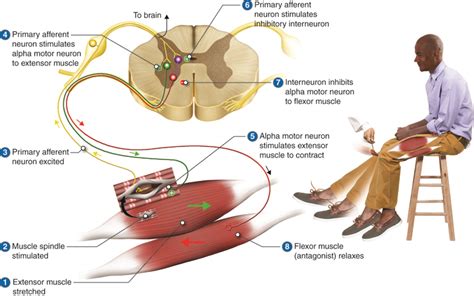
Table of Contents
Reflexes That Control Skeletal Muscle Are Called Somatic Reflexes
Reflexes are involuntary and nearly instantaneous movements in response to a stimulus. They're crucial for our survival, allowing us to react quickly to potentially harmful situations without conscious thought. But what about the specific reflexes that govern the movement of our skeletal muscles, the muscles responsible for our voluntary movements? The answer is somatic reflexes. This comprehensive guide will delve deep into the world of somatic reflexes, exploring their mechanisms, classifications, common examples, and clinical significance.
Understanding Somatic Reflexes: A Deep Dive
Somatic reflexes are involuntary, stereotyped, and rapid responses mediated by the somatic nervous system. Unlike autonomic reflexes, which regulate the internal organs and glands, somatic reflexes control skeletal muscles. This means they are responsible for movements like withdrawing your hand from a hot stove or your knee jerking when tapped.
The pathway a somatic reflex follows is known as a reflex arc. This arc comprises several key components:
1. The Receptor: Sensing the Stimulus
The reflex arc begins with a receptor, a specialized sensory neuron that detects a specific stimulus. This could be a mechanoreceptor (detecting pressure or stretch), a thermoreceptor (detecting temperature changes), or a nociceptor (detecting pain). For example, in the knee-jerk reflex, the receptor is a muscle spindle within the quadriceps muscle, sensitive to stretch.
2. The Sensory Neuron: Transmitting the Signal
The receptor is connected to a sensory neuron (also called an afferent neuron), which transmits the sensory information to the central nervous system (CNS). This signal is typically an action potential, a rapid electrical signal that travels along the axon of the sensory neuron. The sensory neuron's axon enters the spinal cord through the dorsal root.
3. The Integration Center: Processing the Information
The sensory neuron synapses with one or more interneurons within the CNS (typically the spinal cord, but sometimes the brainstem). The interneurons act as processing units, integrating information from multiple sensory neurons and determining the appropriate motor response. In some simpler reflexes, the sensory neuron may directly synapse with the motor neuron, bypassing the interneuron altogether. This integration center is where the decision to initiate a reflex is made.
4. The Motor Neuron: Initiating the Response
The integrated signal is then transmitted from the interneuron (or directly from the sensory neuron) to a motor neuron (also called an efferent neuron). The motor neuron carries the signal from the CNS to the effector organ, in this case, the skeletal muscle.
5. The Effector: Producing the Movement
The effector, the skeletal muscle, receives the signal from the motor neuron and contracts, producing the reflexive movement. This contraction is the observable outcome of the reflex arc. For instance, in the knee-jerk reflex, the contraction of the quadriceps muscle causes the leg to extend.
Classifying Somatic Reflexes
Somatic reflexes can be classified in several ways, depending on the factors being considered:
1. Based on the Number of Synapses:
-
Monosynaptic reflexes: These reflexes involve only one synapse between the sensory neuron and the motor neuron. The classic example is the stretch reflex, like the knee-jerk reflex. The speed and simplicity of monosynaptic reflexes make them crucial for maintaining posture and balance.
-
Polysynaptic reflexes: These reflexes involve one or more interneurons between the sensory neuron and the motor neuron. This allows for more complex processing and integration of information, enabling more nuanced and coordinated responses. Examples include the withdrawal reflex (pulling your hand away from a hot object) and the crossed-extensor reflex (extending the opposite leg to maintain balance when withdrawing a leg).
2. Based on the Type of Stimulus:
-
Stretch reflexes: These reflexes are triggered by the stretching of a muscle. Muscle spindles within the muscle detect the stretch and initiate a reflex contraction of the same muscle. This helps maintain muscle tone and prevent overstretching.
-
Withdrawal reflexes: These reflexes are elicited by noxious stimuli, such as pain or heat. They involve the rapid withdrawal of a limb or body part from the harmful stimulus.
-
Other reflexes: There are numerous other reflexes triggered by various stimuli, including light (pupillary reflex), sound (acoustic reflex), and touch (cutaneous reflexes).
Common Examples of Somatic Reflexes
Several common somatic reflexes are frequently examined in clinical settings to assess the integrity of the nervous system. Let's look at some prominent examples:
1. The Stretch Reflex (Myotatic Reflex): The Knee-Jerk Reflex
The classic example of a monosynaptic reflex is the knee-jerk reflex, also known as the patellar reflex. Tapping the patellar tendon just below the kneecap stretches the quadriceps muscle. This stretch activates muscle spindles within the quadriceps, triggering sensory neurons that directly synapse with motor neurons innervating the quadriceps. The resultant contraction of the quadriceps extends the leg. Simultaneously, an inhibitory interneuron inhibits the antagonistic hamstring muscle, ensuring smooth and efficient leg extension. Absence or hyperactivity of this reflex can indicate neurological problems.
2. The Withdrawal Reflex (Flexor Reflex): Pulling Away from Pain
The withdrawal reflex is a polysynaptic reflex triggered by noxious stimuli like pain or extreme heat. For example, touching a hot stove activates nociceptors in the skin. These nociceptors send signals to the spinal cord via sensory neurons, which synapse with interneurons. These interneurons then activate motor neurons innervating the flexor muscles in the arm, causing the arm to withdraw rapidly from the hot surface. This reflex protects the body from further harm.
3. The Crossed-Extensor Reflex: Maintaining Balance
Often accompanying the withdrawal reflex is the crossed-extensor reflex. When you withdraw one leg from a painful stimulus, the crossed-extensor reflex simultaneously extends the opposite leg. This helps maintain balance and prevents you from falling. The sensory neuron from the stimulated leg sends collaterals to the opposite side of the spinal cord, exciting extensor motor neurons and inhibiting flexor motor neurons in the opposite leg.
4. The Plantar Reflex (Babinski Sign): Assessing Neurological Function
The plantar reflex involves stroking the sole of the foot. In adults, this typically causes plantar flexion (curling of the toes). However, in infants and individuals with certain neurological conditions, the response is dorsiflexion (extension of the big toe) and fanning of the other toes, known as the Babinski sign. The Babinski sign indicates damage to the corticospinal tract, a pathway crucial for voluntary motor control.
Clinical Significance of Somatic Reflexes
Assessing somatic reflexes is a crucial component of neurological examinations. Abnormal reflexes can provide valuable information about the health of the nervous system and can help diagnose various neurological conditions. Here are some ways somatic reflexes are used clinically:
-
Identifying Neurological Lesions: Changes in reflex responses, such as hyperreflexia (exaggerated reflexes), hyporeflexia (diminished reflexes), or the presence of abnormal reflexes like the Babinski sign, can pinpoint lesions in specific parts of the nervous system, including the spinal cord, brainstem, or brain.
-
Monitoring Neurological Disease Progression: Tracking changes in reflex responses over time can help monitor the progression of neurological diseases such as multiple sclerosis, amyotrophic lateral sclerosis (ALS), and Guillain-Barré syndrome.
-
Assessing Spinal Cord Injuries: Reflex tests are essential in evaluating the extent and location of spinal cord injuries. Specific reflex patterns can help determine the level of spinal cord damage.
-
Evaluating Drug Effects: Certain medications can affect the nervous system and alter reflex responses. Monitoring reflexes can help assess the effects of drugs on the nervous system.
-
Diagnosing Peripheral Neuropathies: Peripheral neuropathies, conditions affecting the peripheral nerves, often manifest as altered reflexes. Reflex testing can help diagnose and monitor these conditions.
Conclusion: The Importance of Somatic Reflexes
Somatic reflexes are essential for our survival and well-being. These rapid, involuntary movements protect us from harm and enable us to maintain posture and balance. Understanding the mechanisms, classifications, and clinical significance of somatic reflexes is crucial for healthcare professionals and anyone interested in the workings of the human nervous system. The detailed examination of these reflexes provides invaluable diagnostic insights into the health and integrity of the nervous system, underscoring their importance in clinical practice and neurological research. Further investigation into specific somatic reflexes, including variations based on age, genetics, and individual differences, promises to yield a deeper understanding of human physiology and potential therapeutic interventions for neurological disorders.
Latest Posts
Latest Posts
-
Amoeba Sisters Video Recap Answer Key Cell Transport
Apr 02, 2025
-
What Does Extension Of A Point Refer To
Apr 02, 2025
-
Decidimos Venir A Este Restaurante Porque Mi Jefe
Apr 02, 2025
-
Skills Module 3 0 Intravenous Medication Administration Posttest
Apr 02, 2025
-
America The Story Of Us Episode 9 Bust
Apr 02, 2025
Related Post
Thank you for visiting our website which covers about Reflexes That Control Skeletal Muscle Are Called __________. . We hope the information provided has been useful to you. Feel free to contact us if you have any questions or need further assistance. See you next time and don't miss to bookmark.
