Skeletal Muscle Cells Are Grouped Into Bundles Called __________.
Breaking News Today
Apr 05, 2025 · 6 min read

Table of Contents
Skeletal Muscle Cells are Grouped into Bundles Called Fascicles
Skeletal muscle tissue, the type of muscle responsible for voluntary movement, isn't a homogenous mass. Instead, it's a highly organized structure composed of numerous components working in concert. A crucial aspect of this organization involves the grouping of muscle cells, also known as muscle fibers, into bundles. These bundles are called fascicles. Understanding fascicle arrangement is key to comprehending muscle function, strength, and overall anatomy.
The Hierarchical Structure of Skeletal Muscle
To fully appreciate the role of fascicles, it's essential to understand the hierarchical structure of skeletal muscle:
1. Muscle Fiber (Muscle Cell):
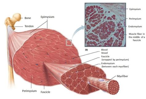
The basic unit of skeletal muscle is the muscle fiber, a long, cylindrical multinucleated cell. Each fiber contains numerous myofibrils, the contractile units responsible for muscle contraction. These myofibrils are arranged in a highly organized pattern of sarcomeres, the smallest functional units of muscle contraction. The sarcomeres contain the protein filaments actin and myosin, which slide past each other during contraction, causing the muscle fiber to shorten.
2. Endomysium:
Surrounding each individual muscle fiber is a delicate layer of connective tissue called the endomysium. This thin sheath provides support and insulation for each fiber, allowing for efficient transmission of nerve impulses and nutrient delivery. It also contains capillaries and nerve fibers essential for muscle function.
3. Fascicle:
Multiple muscle fibers are bundled together to form a fascicle. This is where the answer to our question lies. Fascicles are not just random collections of fibers; their arrangement is highly organized and contributes significantly to the overall shape and function of the muscle. The connective tissue surrounding each fascicle is called the perimysium.
4. Perimysium:
The perimysium is a thicker layer of connective tissue than the endomysium, encasing the fascicles. It provides structural support to the fascicles and allows for the transmission of blood vessels and nerves to the individual fibers within the fascicle. The perimysium also plays a crucial role in transmitting force generated by the muscle fibers.
5. Epimysium:
Several fascicles are bundled together to form the entire muscle, which is encased in a dense layer of connective tissue called the epimysium. This outermost layer provides overall structural integrity to the muscle and connects the muscle to its tendons, which attach the muscle to bones.
6. Tendon:
The epimysium extends beyond the muscle belly to form the tendon. Tendons are strong, fibrous cords that transmit the force generated by the muscle to the bones, allowing for movement.
Fascicle Arrangement and Muscle Function
The arrangement of fascicles within a muscle significantly influences its overall function. Different arrangements result in muscles with varying capabilities in terms of power, range of motion, and the direction of force generated. Here are some common fascicle arrangements:
1. Parallel Fascicle Arrangement:
In muscles with a parallel fascicle arrangement, the fascicles run parallel to the long axis of the muscle. These muscles tend to have a greater range of motion but relatively less power compared to other arrangements. Examples include the rectus abdominis and sartorius muscles.
Advantages of Parallel Arrangement:
- Greater Range of Motion: Due to the parallel arrangement, these muscles can lengthen and shorten significantly.
- High Speed of Contraction: The long fibers allow for rapid contraction.
Disadvantages of Parallel Arrangement:
- Lower Power: Compared to pennate muscles, they have less power.
- Smaller Cross-sectional Area: Results in less overall force generation.
2. Convergent Fascicle Arrangement:
In convergent muscles, the fascicles converge toward a single tendon from a broad origin. This arrangement allows for force generation from a wide area to be focused onto a smaller insertion point. The pectoralis major is a classic example of a convergent muscle.
Advantages of Convergent Arrangement:
- Force Focusing: The converging fascicles focus force onto a smaller area for powerful movements.
- Versatile Movement: The arrangement allows for a wider range of movements due to the variations in fiber orientation.
Disadvantages of Convergent Arrangement:
- Reduced Power in Specific Directions: Because of the varying fiber orientation, force generation may not be as efficient in every direction.
3. Pennate Fascicle Arrangement:
Pennate muscles have fascicles that attach obliquely (at an angle) to a central tendon that runs the length of the muscle. This arrangement results in a greater number of muscle fibers packed into a given volume, increasing the overall power of the muscle. There are three subtypes:
- Unipennate: Fascicles attach to only one side of the tendon (e.g., extensor digitorum longus).
- Bipennate: Fascicles attach to both sides of the central tendon (e.g., rectus femoris).
- Multipennate: Fascicles attach to multiple tendons, creating a complex arrangement (e.g., deltoid).
Advantages of Pennate Arrangement:
- High Power: The oblique arrangement of fibers allows for a greater number of fibers to be packed into a smaller space, resulting in greater power.
- High Force Generation: The oblique pull amplifies force generated.
Disadvantages of Pennate Arrangement:
- Reduced Range of Motion: The oblique arrangement limits the range of motion compared to parallel muscles.
- Slower Contraction Speed: The shorter muscle fibers lead to slower contraction speeds.
4. Circular Fascicle Arrangement:
Circular fascicles are arranged in concentric rings, surrounding an opening or orifice. These muscles act as sphincters, closing or constricting the opening. The orbicularis oculi (muscle around the eye) and orbicularis oris (muscle around the mouth) are examples of circular muscles.
Advantages of Circular Arrangement:
- Precise Control of Openings: These muscles are ideal for controlling the opening and closing of orifices.
Disadvantages of Circular Arrangement:
- Limited Movement Range: Their function is restricted to constriction or dilation.
5. Spiral Fascicle Arrangement:
In spiral muscles, fascicles run obliquely around the long axis of the muscle. This arrangement allows for rotation and other complex movements. The supinator muscle in the forearm is an example of a spiral muscle.
Advantages of Spiral Arrangement:
- Rotation and Complex Movements: The spiral organization facilitates rotational and other nuanced movements.
Disadvantages of Spiral Arrangement:
- Complex Biomechanics: The mechanics of force transmission can be more complex to analyze.
Clinical Significance of Fascicle Arrangement
Understanding fascicle arrangement is crucial in several clinical contexts:
- Muscle Injury Diagnosis: The pattern of muscle damage can be related to the fascicle arrangement and can inform diagnosis and treatment.
- Rehabilitation: Knowing fascicle arrangement is essential for designing effective rehabilitation programs following muscle injuries.
- Surgical Procedures: Surgeons must consider fascicle arrangement when performing procedures involving skeletal muscles.
- Sports Medicine: Understanding muscle architecture, including fascicle arrangement, is essential for optimizing athletic performance and injury prevention.
Conclusion
The arrangement of muscle fibers into fascicles is a critical feature of skeletal muscle architecture. The various fascicle arrangements—parallel, convergent, pennate, circular, and spiral—contribute significantly to the diverse functional capabilities of muscles throughout the body. Understanding this hierarchical organization and the resulting functional variations is fundamental to comprehending muscle physiology, biomechanics, and clinical applications. The detailed study of fascicle architecture continues to reveal new insights into muscle function and disease processes, making it a vibrant area of ongoing research. The next time you think about movement, remember the intricate network of fascicles working tirelessly to power your every action.
Latest Posts
Latest Posts
-
What Does Being A Manager Offer To An Employee
Apr 05, 2025
-
Ati Dosage Calculation Proctored Exam 35 Questions
Apr 05, 2025
-
Disorderly Conduct Is An Example Of A Crime Against
Apr 05, 2025
-
Gramatica C Subject Pronouns And Ser Page 6
Apr 05, 2025
-
A Check That A Bank Refuses To Pay
Apr 05, 2025
Related Post
Thank you for visiting our website which covers about Skeletal Muscle Cells Are Grouped Into Bundles Called __________. . We hope the information provided has been useful to you. Feel free to contact us if you have any questions or need further assistance. See you next time and don't miss to bookmark.
