This Figure Depicts The Basic Anatomy Of The __________.
Breaking News Today
Mar 16, 2025 · 7 min read
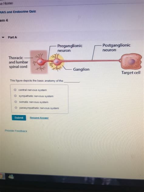
Table of Contents
This Figure Depicts the Basic Anatomy of the Human Heart
This detailed article will explore the intricate anatomy of the human heart, delving into its chambers, valves, vessels, and the electrical conduction system that orchestrates its rhythmic beating. We'll also touch upon the heart's location within the thoracic cavity and its relationship with surrounding structures. Understanding the heart's anatomy is crucial for comprehending its function and the various cardiovascular diseases that can affect it. This comprehensive guide is designed to be informative and accessible, catering to both students and anyone with a general interest in human anatomy and physiology.
Introduction: The Heart – A Marvel of Biological Engineering
The human heart, a fist-sized muscular organ, is the powerhouse of the circulatory system. Its tireless work of pumping blood throughout the body is essential for life. This organ isn't just a simple pump; it's a sophisticated, self-regulating machine with a complex internal structure that enables it to efficiently deliver oxygen and nutrients while removing waste products. Understanding its anatomy is key to grasping its incredible functionality.
1. Chambers of the Heart: Four Compartments, Four Crucial Roles
The heart is divided into four chambers: two atria (singular: atrium) and two ventricles. These chambers work in a coordinated manner to ensure unidirectional blood flow.
-
Right Atrium: This chamber receives deoxygenated blood returning from the body via the superior and inferior vena cava. The superior vena cava brings blood from the upper body, while the inferior vena cava carries blood from the lower body.
-
Right Ventricle: The right atrium pumps the deoxygenated blood into the right ventricle. From here, the blood is pumped through the pulmonary valve into the pulmonary artery, which carries it to the lungs for oxygenation.
-
Left Atrium: After oxygenation in the lungs, the now-oxygenated blood returns to the heart via the pulmonary veins and enters the left atrium.
-
Left Ventricle: The left atrium pumps the oxygenated blood into the left ventricle. This is the heart's most powerful chamber; it pumps the oxygenated blood through the aortic valve into the aorta, the body's largest artery, distributing oxygenated blood throughout the systemic circulation.
2. Heart Valves: Ensuring One-Way Traffic
The heart's valves are critical for maintaining unidirectional blood flow. These valves open and close passively, responding to pressure changes within the chambers.
-
Tricuspid Valve: Located between the right atrium and the right ventricle, this valve has three leaflets (cusps) that prevent backflow of blood from the ventricle into the atrium.
-
Pulmonary Valve: Situated at the exit of the right ventricle, this semilunar valve (with three half-moon-shaped cusps) prevents backflow of blood from the pulmonary artery into the right ventricle.
-
Mitral Valve (Bicuspid Valve): Found between the left atrium and the left ventricle, this valve has two leaflets and prevents backflow from the ventricle into the atrium.
-
Aortic Valve: Located at the exit of the left ventricle, this semilunar valve prevents backflow of blood from the aorta into the left ventricle.
3. Major Blood Vessels: The Highways of the Circulatory System
Several major blood vessels connect the heart to the rest of the circulatory system.
-
Superior and Inferior Vena Cava: These large veins return deoxygenated blood from the upper and lower body, respectively, to the right atrium.
-
Pulmonary Artery: This artery carries deoxygenated blood from the right ventricle to the lungs. Note that this is the only artery in the body carrying deoxygenated blood.
-
Pulmonary Veins: These veins return oxygenated blood from the lungs to the left atrium. These are the only veins in the body carrying oxygenated blood.
-
Aorta: This is the largest artery in the body, carrying oxygenated blood from the left ventricle to the rest of the body. It branches into numerous smaller arteries that supply blood to specific organs and tissues.
4. The Cardiac Conduction System: The Heart's Internal Pacemaker
The rhythmic beating of the heart is not simply a mechanical process; it's orchestrated by a specialized electrical conduction system. This system generates and conducts electrical impulses that stimulate the heart muscle to contract.
-
Sinoatrial (SA) Node: Often called the heart's natural pacemaker, the SA node is located in the right atrium. It generates electrical impulses that initiate each heartbeat.
-
Atrioventricular (AV) Node: Located between the atria and ventricles, the AV node delays the electrical impulse, allowing the atria to fully contract before the ventricles begin to contract.
-
Bundle of His: This bundle of specialized conducting fibers transmits the impulse from the AV node to the ventricles.
-
Purkinje Fibers: These fibers rapidly distribute the electrical impulse throughout the ventricles, causing them to contract simultaneously and efficiently.
5. Heart Location and Surrounding Structures:
The heart is situated within the mediastinum, the central compartment of the thoracic cavity. It's positioned slightly to the left of the midline, between the lungs. The heart is surrounded by a double-layered sac called the pericardium, which protects it and helps to lubricate its movement. The pericardium consists of the fibrous pericardium (outer layer) and the serous pericardium (inner layer).
6. Heart Wall Layers: Structure and Function
The heart wall itself comprises three distinct layers:
-
Epicardium: The outermost layer, also known as the visceral pericardium, is a thin serous membrane that covers the heart's surface.
-
Myocardium: This is the thickest layer and consists of cardiac muscle tissue. The myocardium is responsible for the heart's powerful contractions. The thickness of the myocardium varies depending on the chamber; the left ventricle has the thickest myocardium due to its role in pumping blood throughout the systemic circulation.
-
Endocardium: The innermost layer, lining the heart chambers and valves, is a thin, smooth endothelium that minimizes friction during blood flow.
7. Coronary Circulation: Nourishing the Heart Muscle
While the heart pumps blood to the rest of the body, it requires its own blood supply to function. This is provided by the coronary arteries, which branch off from the aorta just as it leaves the left ventricle. These arteries deliver oxygenated blood to the heart muscle, while the coronary veins return deoxygenated blood back to the right atrium. The efficiency of coronary circulation is critical for the heart's health and performance. Blockages or narrowing in the coronary arteries can lead to coronary artery disease (CAD) and potentially heart attacks.
8. Clinical Significance: Understanding Heart Conditions
Understanding the heart's anatomy is fundamental to diagnosing and treating various cardiovascular diseases. Many conditions, including congenital heart defects, valve disorders (e.g., mitral valve prolapse, aortic stenosis), coronary artery disease (CAD), arrhythmias, and heart failure, directly relate to the structure and function of the heart's different components. For example, a defect in the septums (the walls separating the heart chambers) can lead to a mixing of oxygenated and deoxygenated blood, reducing the efficiency of oxygen delivery to the body.
9. Advanced Anatomical Considerations:
Beyond the basic anatomy outlined above, a deeper understanding of the heart involves investigating the intricate microanatomy of cardiac muscle cells, the precise arrangement of myocardial fibers, and the neurovascular supply that supports the heart's intricate network. Advanced imaging techniques, such as echocardiography, cardiac MRI, and cardiac CT scans, allow for detailed visualization of the heart's internal structures and help diagnose various conditions.
10. Conclusion: The Heart's Enduring Importance
The human heart, with its complex interplay of chambers, valves, vessels, and the electrical conduction system, is a testament to the remarkable engineering of the human body. A thorough understanding of its anatomy is paramount for medical professionals and is valuable for anyone seeking to appreciate the intricate workings of this vital organ. Continuous research into cardiac anatomy and physiology is vital for developing innovative treatments and preventative strategies for cardiovascular diseases, which remain a leading cause of mortality worldwide. By appreciating the complexity and significance of the heart's structure, we can better understand its importance in maintaining life and overall health. The detailed exploration of this incredible organ underscores the ongoing necessity for advancements in cardiovascular research and care.
Latest Posts
Latest Posts
-
The Direct Carry Is Used To Transfer A Patient
Mar 18, 2025
-
The Emancipation Proclamation Of January 1 1863 Quizlet
Mar 18, 2025
-
These Cards Will Get You Drunk Quizlet
Mar 18, 2025
-
Did Quizlet Get Rid Of Q Chat
Mar 18, 2025
-
Myasthenia Gravis Is An Autoimmune Disease In Which Quizlet
Mar 18, 2025
Related Post
Thank you for visiting our website which covers about This Figure Depicts The Basic Anatomy Of The __________. . We hope the information provided has been useful to you. Feel free to contact us if you have any questions or need further assistance. See you next time and don't miss to bookmark.
