Correctly Label The Anterior Muscles Of The Thigh
Breaking News Today
Mar 15, 2025 · 6 min read
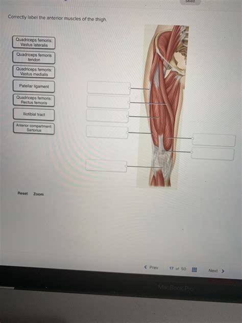
Table of Contents
Correctly Labeling the Anterior Muscles of the Thigh: A Comprehensive Guide
Understanding the anterior muscles of the thigh is crucial for anyone studying anatomy, kinesiology, or involved in fields like physical therapy, athletic training, or fitness. This region is complex, housing several muscles with distinct functions, origins, and insertions. Accurate labeling is paramount for effective communication and understanding of movement and potential injuries. This comprehensive guide will break down the anterior thigh muscles, providing detailed descriptions and helpful tips for accurate identification.
The Key Players: A Detailed Look at Each Muscle
The anterior compartment of the thigh primarily contains muscles responsible for hip flexion and knee extension. Let's explore each muscle individually:
1. Sartorius: The "Tailor's Muscle"
- Origin: Anterior superior iliac spine (ASIS)
- Insertion: Proximal, medial shaft of the tibia (pes anserinus)
- Action: Hip flexion, abduction, and lateral rotation; knee flexion
The sartorius is the longest muscle in the human body. Its name, derived from the Latin word "sartor" (tailor), reflects its action of externally rotating the hip, a position historically used by tailors when sitting cross-legged. It plays a significant role in hip and knee movements, contributing to a wide range of actions. Remember its unique, long, strap-like appearance. This will help you distinguish it from other muscles in the region.
2. Quadriceps Femoris: The Powerhouse of Knee Extension
The quadriceps femoris isn't a single muscle but a group of four muscles: rectus femoris, vastus lateralis, vastus medialis, and vastus intermedius. Understanding each component is vital for accurate labeling.
2.1 Rectus Femoris: The Only Bi-articular Muscle
- Origin: Anterior inferior iliac spine (AIIS) and superior acetabulum
- Insertion: Tibial tuberosity via the patellar tendon
- Action: Hip flexion and knee extension
The rectus femoris is unique among the quadriceps as it crosses both the hip and knee joints, making it a bi-articular muscle. This means it acts on both joints simultaneously. Look for its superficial position and its relatively straight course down the thigh. Its origin at the pelvis is a key distinguishing feature.
2.2 Vastus Lateralis: The Largest of the Quadriceps
- Origin: Greater trochanter, intertrochanteric line, linea aspera, and lateral supracondylar line of the femur
- Insertion: Tibial tuberosity via the patellar tendon
- Action: Knee extension
The vastus lateralis is the largest of the quadriceps muscles. Its name reflects its position – it's located on the lateral side of the thigh. Observe its substantial size and its origin along the lateral aspect of the femur. It plays a dominant role in powerful knee extension movements.
2.3 Vastus Medialis: Stabilizing the Knee
- Origin: Intertrochanteric line, medial supracondylar line, and medial aspect of the femur
- Insertion: Tibial tuberosity via the patellar tendon
- Action: Knee extension
The vastus medialis is located on the medial side of the thigh. It's crucial for stabilizing the patella (kneecap) during knee extension. Note its position and its often slightly feathered appearance. This muscle's proper function is essential for preventing patellar tracking issues.
2.4 Vastus Intermedius: Deep and Often Overlooked
- Origin: Anterior and lateral surfaces of the femur
- Insertion: Tibial tuberosity via the patellar tendon
- Action: Knee extension
The vastus intermedius lies deep to the rectus femoris, making it harder to visualize. It plays a significant role in knee extension, but its deep position often makes it challenging to identify. Remember its deep location and its relationship to the rectus femoris. Understanding its location requires a deeper understanding of the layers of the anterior thigh.
Tips for Accurate Labeling: A Practical Approach
Accurately labeling the anterior thigh muscles requires careful observation and a systematic approach. Here are some practical tips to enhance your understanding and labeling accuracy:
- Start with the Superficial Muscles: Begin by identifying the more superficial muscles, such as the sartorius and rectus femoris. Their prominent positions make them easier to locate.
- Utilize Anatomical Landmarks: Use readily identifiable bony landmarks like the ASIS, AIIS, and the greater trochanter to guide your muscle identification. These landmarks provide crucial reference points.
- Trace Muscle Fibers: Carefully trace the direction and pattern of muscle fibers. This helps determine the muscle's origin and insertion, aiding in accurate identification.
- Consider Muscle Actions: Thinking about the muscle’s actions can provide additional clues. For example, the hip flexion action of the rectus femoris helps differentiate it from the other quadriceps muscles.
- Use Anatomical Models and Diagrams: Utilize anatomical models and diagrams to supplement your knowledge and clarify any uncertainties. Visual aids are invaluable learning tools.
- Practice, Practice, Practice: Consistent practice with anatomical models, diagrams, and real-life observation is crucial for mastering muscle identification. Repetition solidifies your understanding.
- Understand the Relationships Between Muscles: Pay attention to the relationships between different muscles. Understanding how muscles interact and overlap contributes to accurate labeling. For instance, the vastus intermedius lies beneath the rectus femoris.
Clinical Significance: Why Accurate Labeling Matters
Accurate labeling of the anterior thigh muscles is crucial for various clinical applications:
- Diagnosis of Injuries: Precise identification of injured muscles is vital for appropriate diagnosis and treatment. Mislabeling can lead to delayed or ineffective treatment.
- Physical Therapy and Rehabilitation: Accurate muscle identification enables targeted exercises and rehabilitation strategies for optimal recovery.
- Surgical Procedures: Accurate anatomical knowledge is paramount during surgical procedures to minimize complications and maximize success.
- Sports Medicine: Understanding the function and potential vulnerabilities of these muscles is crucial for injury prevention and effective rehabilitation in athletes.
Common Errors and How to Avoid Them
Several common errors occur when labeling anterior thigh muscles:
- Confusing Rectus Femoris with Sartorius: These muscles share some similar actions, potentially leading to confusion. Remember, the sartorius is much longer and more superficial, with a more lateral course.
- Misidentifying Vastus Medialis and Vastus Lateralis: Their positions are opposite. Carefully note the medial and lateral aspects of the thigh.
- Overlooking the Vastus Intermedius: Its deep position makes it easily overlooked. Remember to consider the deeper layers of muscle.
- Incorrectly Identifying Muscle Origins and Insertions: Pay close attention to the attachments of each muscle.
Conclusion: Mastering the Anterior Thigh
Mastering the accurate labeling of the anterior thigh muscles requires a dedicated approach. By employing the strategies outlined in this comprehensive guide, combining meticulous observation with a strong theoretical understanding, you can confidently identify and label the sartorius and the four muscles of the quadriceps femoris group. Remember, consistent practice and the use of various learning resources are key to success. This anatomical knowledge is invaluable, not only for academic purposes but also for clinical practice and ensuring accurate communication within the medical and fitness fields. The detailed understanding of these muscles is fundamental to effective diagnosis, treatment, and rehabilitation. Remember to always approach anatomical study with diligence and a keen eye for detail.
Latest Posts
Latest Posts
-
What Is The Best Definition Of Marginal Revenue Quizlet
Mar 18, 2025
-
The Superficial Temporal Artery Can Be Palpated Quizlet
Mar 18, 2025
-
Rn Comprehensive Online Practice 2023 B With Ngn Quizlet
Mar 18, 2025
-
Administrative Civil Or Criminal Sanctions Cui Quizlet
Mar 18, 2025
-
Sinners In The Hands Of An Angry God Quizlet
Mar 18, 2025
Related Post
Thank you for visiting our website which covers about Correctly Label The Anterior Muscles Of The Thigh . We hope the information provided has been useful to you. Feel free to contact us if you have any questions or need further assistance. See you next time and don't miss to bookmark.
