The Superficial Temporal Artery Can Be Palpated Quizlet
Breaking News Today
Mar 18, 2025 · 5 min read
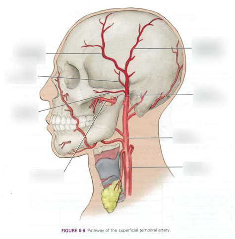
Table of Contents
The Superficial Temporal Artery: Palpation, Anatomy, and Clinical Significance
The superficial temporal artery (STA) is a readily palpable artery located on the side of the head, making it a crucial landmark for both anatomical study and clinical practice. Its accessibility and consistent location make it ideal for various procedures, including blood pressure measurement and temporal artery biopsy. This article delves into the detailed anatomy of the STA, its palpation technique, and its clinical significance, clarifying common misconceptions and providing a comprehensive understanding of this important vessel.
Anatomy of the Superficial Temporal Artery
The STA is a terminal branch of the external carotid artery (ECA), one of the two major branches of the common carotid artery. Understanding the ECA's branching pattern is crucial for comprehending the STA's origin and course. The ECA ascends in the neck, branching into several arteries before terminating as the STA and the maxillary artery.
Origin and Course:
The STA originates posterior to the neck of the mandible, deep within the parotid gland. After exiting the parotid gland, it becomes superficial, running superiorly and anteriorly across the zygomatic arch. It then ascends across the temporal fossa, lying superficial to the temporalis muscle. The artery's course can be slightly variable, but its general trajectory remains relatively consistent.
Branches and Distribution:
Along its course, the STA gives off several smaller branches:
- Anterior auricular branch: Supplies blood to the anterior part of the auricle (outer ear).
- Zygomatico-orbital branch: Provides blood to the tissues around the zygomatic arch and orbit.
- Middle temporal artery: Supplies blood to the temporal region.
- Parietal branch: Supplies blood to the parietal region of the scalp.
These branches contribute to the rich vascular supply of the scalp and surrounding structures. The STA's extensive branching network ensures adequate perfusion even in cases of partial occlusion.
Relationship with other structures:
The STA's superficial location means it's closely associated with various anatomical structures. Understanding these relationships is crucial for accurate palpation and for avoiding inadvertent injury during procedures:
- Temporalis Muscle: The STA lies superficial to the temporalis muscle, making the muscle a palpable landmark during artery location.
- Auricularis Anterior Muscle: This small muscle, situated anterior to the ear, provides another surface marker that assists in locating the STA.
- Parotid Gland: The STA emerges from the parotid gland; its location relative to this gland is important in surgical procedures.
- Skin and Subcutaneous Tissue: The artery is very superficial, lying just beneath the skin and subcutaneous tissue. This proximity facilitates easy palpation.
- Cranial Nerves: While not directly in contact, the STA's course runs in close proximity to certain cranial nerves, raising the possibility of accidental injury during surgical interventions.
Palpating the Superficial Temporal Artery: A Step-by-Step Guide
Palpating the STA is a relatively straightforward procedure, but proper technique ensures accurate identification and avoids undue pressure.
1. Patient Positioning:
The patient should be seated comfortably with their head slightly tilted. This improves accessibility and visualization of the temporal region.
2. Landmark Identification:
Begin by locating the zygomatic arch, the bony structure that forms the prominence of the cheek. The STA lies superior and anterior to the arch.
3. Palpation Technique:
Use the pads of your index and middle fingers to gently palpate the temporal region, moving superiorly from the zygomatic arch towards the hairline. Avoid excessive pressure, as this can cause discomfort and potentially obscure the artery's pulse.
4. Pulse Detection:
The STA's pulse is usually readily detectable as a rhythmic pulsation. The pulsation is typically stronger when the patient is under stress or exertion. The pulsation should feel relatively superficial and distinct.
5. Confirmation:
Once a pulse is detected, carefully trace the artery's course to confirm its identity. The STA's path is generally consistent, running upwards towards the hairline along the temporal line.
Common Mistakes to Avoid:
- Excessive pressure: Avoid applying too much pressure, as this can obscure the pulse and cause discomfort.
- Incorrect location: Ensure that the pulse is located superior and anterior to the zygomatic arch to differentiate it from other superficial vessels.
- Misidentification: Confusing the STA with other superficial vessels in the area should be avoided. Careful observation of the anatomical location and the pulsatile nature of the vessel is crucial.
Clinical Significance of the Superficial Temporal Artery
The STA's accessibility and consistent location make it invaluable in various clinical settings:
1. Temporal Artery Biopsy:
The STA is the preferred site for temporal artery biopsy, a procedure used to diagnose giant cell arteritis (GCA), a form of vasculitis. The artery's superficial location allows for easy access and minimal invasiveness during the biopsy procedure.
2. Blood Pressure Measurement:
Although less commonly used than brachial artery palpation, the STA can be used for blood pressure measurement, especially in situations where access to the brachial artery is difficult.
3. Surgical Procedures:
The STA's proximity to the scalp and facial structures makes it an important consideration in various surgical procedures, including those involving the temporal region or face. Knowledge of the artery's anatomy helps surgeons avoid inadvertent injury.
4. Assessment of Vascular Supply:
Palpation of the STA can provide valuable information about the patient's peripheral vascular health. Absent or diminished pulse may indicate a problem with vascular perfusion to the head and neck.
5. Clinical Examination in Neurological Conditions:
The STA's pulsation can be a useful indicator in assessing some neurological conditions, especially in cases involving head injuries or vascular pathologies.
Variations and Anomalies
While the STA's location is typically consistent, anatomical variations can occur. These variations can affect the artery's course, branching pattern, or even its presence. Understanding these possibilities is crucial for accurate diagnosis and treatment. Such variations should be considered during surgical procedures or biopsies to prevent unexpected complications.
Conclusion
The superficial temporal artery is a significant anatomical structure with wide-ranging clinical importance. Its consistent location and superficial position allow for easy palpation, making it a useful landmark in anatomical studies and various medical procedures. The ability to accurately locate and palpate the STA is essential for medical professionals, contributing significantly to accurate diagnosis, minimally invasive procedures, and the overall care of patients. Mastering the technique of STA palpation is not only a skill for medical professionals but also a foundational knowledge point in the study of human anatomy and physiology. Understanding its anatomy, location, and clinical significance can be critical for healthcare providers and medical students alike.
Latest Posts
Latest Posts
-
True Or False Professional And Technical Communication Is Research Oriented
Mar 18, 2025
-
Which Best Describes The Terrorist Planning Cycle
Mar 18, 2025
-
Cdl Combination Test Questions And Answers Pdf
Mar 18, 2025
-
Life Insurance Exam Questions And Answers Pdf
Mar 18, 2025
-
The Direct Carry Is Used To Transfer A Patient
Mar 18, 2025
Related Post
Thank you for visiting our website which covers about The Superficial Temporal Artery Can Be Palpated Quizlet . We hope the information provided has been useful to you. Feel free to contact us if you have any questions or need further assistance. See you next time and don't miss to bookmark.
