Correctly Label The Following Anatomical Features Of The Neuroglia
Breaking News Today
Mar 30, 2025 · 6 min read
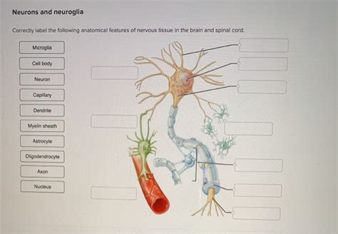
Table of Contents
Correctly Label the Following Anatomical Features of the Neuroglia: A Comprehensive Guide
Neuroglia, also known as glial cells, are non-neuronal cells in the central nervous system (CNS) and the peripheral nervous system (PNS). They are vastly outnumbered by neurons, yet they play crucial roles in supporting and protecting neurons, as well as contributing to overall brain function. Understanding the different types of neuroglia and their specific anatomical features is essential for comprehending the complexities of the nervous system. This comprehensive guide will delve into the key anatomical features of various neuroglia, enabling you to correctly label them with confidence.
Types of Neuroglia and Their Key Features
The neuroglia are diverse, each type exhibiting unique morphological characteristics and functional roles. We'll explore the main types found in the CNS and PNS, emphasizing their distinguishing anatomical features.
Central Nervous System (CNS) Neuroglia
1. Astrocytes: These star-shaped cells are the most abundant glial cells in the CNS. Their extensive processes contact both neurons and blood vessels, forming a crucial link between the two.
-
Key Anatomical Features:
- Stellate shape: Their numerous, radiating processes give them their characteristic star-like appearance.
- Perivascular feet: These specialized processes wrap around blood vessels, forming the blood-brain barrier (BBB). These feet are crucial for regulating the passage of substances between the blood and the brain.
- Intermediate filaments: Astrocytes contain abundant glial fibrillary acidic protein (GFAP) intermediate filaments, providing structural support. These filaments are a key marker for identifying astrocytes.
- Gap junctions: Astrocytes communicate with each other through gap junctions, allowing for rapid signal transmission.
-
Functional Roles: Beyond structural support, astrocytes play critical roles in regulating synaptic transmission, maintaining the extracellular environment, and responding to injury.
2. Oligodendrocytes: These cells are responsible for myelination in the CNS. Unlike Schwann cells in the PNS, a single oligodendrocyte can myelinate multiple axons.
-
Key Anatomical Features:
- Smaller cell body: Compared to astrocytes, oligodendrocytes have smaller cell bodies.
- Fewer processes: They have fewer processes than astrocytes, each process extending to myelinate a segment of an axon.
- Myelin sheath: The myelin sheath, produced by oligodendrocytes, is a lipid-rich insulating layer that increases the speed of nerve impulse conduction. This sheath appears as concentric layers around the axon.
-
Functional Roles: Their primary function is the formation and maintenance of the myelin sheath, essential for efficient neural communication.
3. Microglia: These are the resident immune cells of the CNS, acting as the brain's primary defense against infection and injury.
-
Key Anatomical Features:
- Small, elongated cell body: Microglia have a relatively small, irregular cell body and elongated processes.
- Highly branched processes: Their processes constantly scan the surrounding environment for signs of damage or infection.
- Amoeboid morphology: Upon activation, microglia transform into an amoeboid morphology, becoming more mobile and phagocytic.
-
Functional Roles: They act as phagocytes, engulfing cellular debris, pathogens, and damaged neurons. They also release cytokines, mediating inflammatory responses.
4. Ependymal Cells: These cells line the ventricles of the brain and the central canal of the spinal cord. They are involved in the production and circulation of cerebrospinal fluid (CSF).
-
Key Anatomical Features:
- Cuboidal or columnar shape: Ependymal cells have a cuboidal or columnar shape, forming a continuous epithelial layer.
- Cilia: Many ependymal cells possess cilia on their apical surface, facilitating the movement of CSF.
- Microvilli: Microvilli on their surface increase the surface area for absorption and secretion.
-
Functional Roles: They produce and circulate CSF, providing a protective cushion for the brain and spinal cord. They also play a role in the transport of nutrients and waste products.
Peripheral Nervous System (PNS) Neuroglia
1. Schwann Cells: These cells are the myelinating cells of the PNS. Unlike oligodendrocytes, each Schwann cell myelinates only a single segment of a single axon.
-
Key Anatomical Features:
- Elongated shape: Schwann cells are elongated and wrap around axons.
- Myelin sheath: They produce the myelin sheath in the PNS, similar to oligodendrocytes in the CNS. The myelin sheath is essential for the rapid conduction of nerve impulses.
- Nodes of Ranvier: The gaps between adjacent Schwann cells, known as Nodes of Ranvier, are crucial for saltatory conduction.
-
Functional Roles: Their main function is the myelination of axons in the PNS, ensuring rapid and efficient signal transmission.
2. Satellite Cells: These cells surround neuron cell bodies in ganglia of the PNS, providing structural support and regulating the extracellular environment.
-
Key Anatomical Features:
- Flattened shape: Satellite cells are flattened cells that surround neuron cell bodies.
- Close association with neurons: They form a supportive layer around the neuron cell bodies in ganglia.
-
Functional Roles: They provide structural support and regulate the ionic environment around neuron cell bodies, maintaining homeostasis.
Labeling Practice: A Step-by-Step Approach
To correctly label neuroglial features, follow these steps:
-
Identify the type of neuroglia: First, determine whether the cell is an astrocyte, oligodendrocyte, microglia, ependymal cell, Schwann cell, or satellite cell based on its overall shape and location (CNS or PNS).
-
Focus on characteristic features: Look for distinguishing anatomical features, such as the stellate shape of astrocytes, the myelin sheaths produced by oligodendrocytes and Schwann cells, the highly branched processes of microglia, the cilia on ependymal cells, and the flattened shape of satellite cells.
-
Use specific terminology: Use precise anatomical terms when labeling, such as "perivascular feet" for astrocytes, "Nodes of Ranvier" for myelinated axons, and "myelin sheath" for the insulating layer.
-
Consider the context: The labeling will also depend on the context of the image or diagram. For example, if the image shows a cross-section of a myelinated axon, you should label both the axon and the myelin sheath, indicating whether it is produced by an oligodendrocyte or a Schwann cell.
-
Consult reliable resources: Refer to reputable textbooks, anatomical atlases, and online resources to verify your labeling and deepen your understanding.
Advanced Considerations: Variations and Pathologies
While the descriptions above outline the typical anatomical features of neuroglia, variations exist depending on location, developmental stage, and physiological state. Furthermore, neuroglia are involved in several neurological pathologies. For instance, alterations in astrocyte function have been implicated in neurodegenerative diseases, while microglial activation plays a central role in neuroinflammation. Understanding these complexities enhances the ability to accurately interpret microscopic images and comprehend the intricate interplay between neuroglia and neurological function.
Conclusion: Mastering Neuroglial Anatomy
Correctly labeling the anatomical features of neuroglia requires a thorough understanding of their diverse types, characteristic morphology, and functional roles. By systematically analyzing the visual characteristics and applying appropriate terminology, you can confidently identify and label these crucial components of the nervous system. Remember that consistent study, coupled with the use of high-quality anatomical resources, is key to mastering neuroglial anatomy. This knowledge forms a crucial foundation for understanding the complexities of the brain and nervous system and further research in neuroscience.
Latest Posts
Latest Posts
-
What Is The Total Of Amalas Liabilities
Apr 01, 2025
-
A Patient Who Has Experienced A Back Injury
Apr 01, 2025
-
Define Back Channel Cues List 3 Examples Of Backchannel Cues
Apr 01, 2025
-
How Does Madison Use Comparison To Bolster His Argument
Apr 01, 2025
-
Afferent Signals From External Stimuli Are Carried By The
Apr 01, 2025
Related Post
Thank you for visiting our website which covers about Correctly Label The Following Anatomical Features Of The Neuroglia . We hope the information provided has been useful to you. Feel free to contact us if you have any questions or need further assistance. See you next time and don't miss to bookmark.
