Correctly Label The Following Parts Of The Adrenal Gland.
Breaking News Today
Mar 14, 2025 · 5 min read
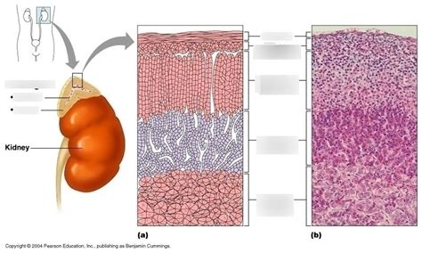
Table of Contents
Correctly Label the Following Parts of the Adrenal Gland: A Comprehensive Guide
The adrenal glands, also known as suprarenal glands, are vital endocrine glands situated atop each kidney. These small, yet powerful organs play a crucial role in regulating numerous bodily functions through the production of various steroid hormones and catecholamines. Understanding the anatomy of the adrenal gland is key to comprehending its complex physiological roles. This comprehensive guide will detail the various parts of the adrenal gland and their corresponding functions, helping you correctly label each component.
The Adrenal Cortex: Layers of Hormonal Power
The adrenal cortex, the outer region of the adrenal gland, comprises three distinct zones, each responsible for synthesizing specific hormones. These zones are arranged in concentric layers, with the outermost layer being the zona glomerulosa, followed by the zona fasciculata, and finally, the innermost zona reticularis.
1. Zona Glomerulosa: The Mineralocorticoid Maestro
The zona glomerulosa, the outermost layer, is characterized by its tightly packed cells arranged in spherical clusters (glomeruli). Its primary function is the production of mineralocorticoids, particularly aldosterone. Aldosterone plays a crucial role in regulating electrolyte balance, primarily sodium and potassium, by influencing their reabsorption in the kidneys. This, in turn, affects blood volume, blood pressure, and overall fluid balance within the body. Dysfunction in this zone can lead to conditions such as Conn's syndrome (primary hyperaldosteronism), characterized by high blood pressure and low potassium levels, or Addison's disease, which involves insufficient aldosterone production.
Keywords: Zona glomerulosa, aldosterone, mineralocorticoids, electrolyte balance, sodium, potassium, blood pressure, Conn's syndrome, Addison's disease, primary hyperaldosteronism.
2. Zona Fasciculata: The Glucocorticoid Giant
The zona fasciculata, the thickest layer of the adrenal cortex, is composed of cells arranged in long cords or fascicles, separated by prominent capillaries. This zone is responsible for the synthesis and secretion of glucocorticoids, with cortisol being the most significant. Cortisol is a crucial hormone involved in numerous metabolic processes:
- Glucose Metabolism: Cortisol promotes gluconeogenesis (the production of glucose from non-carbohydrate sources), ensuring a steady supply of glucose to the body, particularly during periods of stress or fasting.
- Protein Metabolism: It influences protein breakdown in muscles and other tissues, releasing amino acids that can be used for gluconeogenesis or other metabolic needs.
- Fat Metabolism: Cortisol promotes lipolysis (breakdown of fats) in certain areas of the body while promoting lipogenesis (fat storage) in others.
- Immune Response: Cortisol has potent anti-inflammatory and immunosuppressive effects, regulating the immune response and preventing excessive inflammation. This makes it crucial in managing autoimmune diseases and allergic reactions.
- Stress Response: Cortisol plays a central role in the body's response to stress, helping to maintain homeostasis during challenging situations.
Imbalances in cortisol production can lead to Cushing's syndrome (excess cortisol) characterized by weight gain, high blood sugar, and muscle weakness, or Addison's disease (insufficient cortisol), which presents with fatigue, low blood pressure, and salt craving.
Keywords: Zona fasciculata, cortisol, glucocorticoids, glucose metabolism, gluconeogenesis, protein metabolism, fat metabolism, lipolysis, lipogenesis, immune response, anti-inflammatory, immunosuppressive, stress response, Cushing's syndrome, Addison's disease.
3. Zona Reticularis: The Androgen Architect
The innermost layer of the adrenal cortex, the zona reticularis, is characterized by its cells arranged in a network or reticulum. This zone primarily produces adrenal androgens, such as dehydroepiandrosterone (DHEA) and androstenedione. These hormones contribute to the development of secondary sexual characteristics, particularly in women, although their contribution is less significant than that of the gonadal hormones. They also play a role in overall metabolism and energy production. While not as directly involved in immediate life-threatening conditions as the other zones, imbalances in adrenal androgen production can contribute to conditions like hirsutism (excessive hair growth) and virilisation in women.
Keywords: Zona reticularis, adrenal androgens, dehydroepiandrosterone (DHEA), androstenedione, secondary sexual characteristics, hirsutism, virilisation.
The Adrenal Medulla: The Catecholamine Center
The adrenal medulla, the inner region of the adrenal gland, is embryologically derived from neural crest cells and functions as a part of the sympathetic nervous system. It is primarily responsible for the production and secretion of catecholamines, particularly epinephrine (adrenaline) and norepinephrine (noradrenaline). These hormones are crucial for the body's fight-or-flight response:
- Epinephrine: Primarily responsible for increasing heart rate, blood pressure, and blood glucose levels. It also dilates airways, preparing the body for increased physical activity and stress.
- Norepinephrine: Primarily responsible for increasing blood pressure and constricting blood vessels in non-essential organs, diverting blood flow to muscles and vital organs. It also plays a role in alertness and attention.
Dysfunction in the adrenal medulla can lead to conditions such as pheochromocytoma, a rare tumor that causes excessive release of catecholamines, leading to episodes of severe hypertension, palpitations, and headaches.
Keywords: Adrenal medulla, catecholamines, epinephrine (adrenaline), norepinephrine (noradrenaline), fight-or-flight response, heart rate, blood pressure, blood glucose, pheochromocytoma, hypertension, palpitations, headaches.
Clinical Significance and Diagnostic Techniques
Understanding the different parts of the adrenal gland and their hormonal functions is crucial for diagnosing and managing a wide range of endocrine disorders. Various diagnostic techniques are employed to assess adrenal gland function, including:
- Blood tests: Measuring levels of cortisol, aldosterone, and other hormones.
- Urine tests: Analyzing the excretion of hormones and their metabolites.
- Imaging studies: Techniques like CT scans and MRI scans can visualize the adrenal glands and detect abnormalities such as tumors.
- Stimulation and suppression tests: These tests evaluate the responsiveness of the adrenal glands to various stimuli and help determine the cause of hormonal imbalances.
Conclusion: Mastering the Adrenal Gland's Anatomy
The adrenal glands, with their intricate layers and diverse hormonal secretions, are essential for maintaining homeostasis and responding to stress. By correctly labeling the components—the zona glomerulosa, zona fasciculata, zona reticularis, and adrenal medulla—and understanding their respective roles, you gain a fundamental appreciation for the critical function of these vital organs. This knowledge is essential for healthcare professionals and anyone interested in human physiology and endocrinology. Further research into specific diseases and treatments related to adrenal dysfunction will provide a more complete understanding of this fascinating organ system. Remember to always consult a medical professional for any health concerns. This information is intended for educational purposes only and should not be considered medical advice.
Latest Posts
Latest Posts
-
Dna Is Made Up Of Repeating Units Called
Mar 14, 2025
-
Where Are The Sensors For The Arterial Baroreceptor Reflex Located
Mar 14, 2025
-
Correctly Label The Following Parts Of A Renal Corpuscle
Mar 14, 2025
-
Correctly Label The Following Anatomical Parts Of A Kidney
Mar 14, 2025
-
Using Social Media To Support Activities Such As Producing Maps
Mar 14, 2025
Related Post
Thank you for visiting our website which covers about Correctly Label The Following Parts Of The Adrenal Gland. . We hope the information provided has been useful to you. Feel free to contact us if you have any questions or need further assistance. See you next time and don't miss to bookmark.
