Label The Integumentary Structures And Areas Indicated In The Diagram
Breaking News Today
Mar 28, 2025 · 6 min read
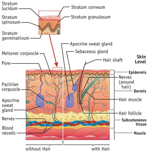
Table of Contents
Label the Integumentary Structures and Areas Indicated in the Diagram: A Comprehensive Guide
The integumentary system, our body's largest organ system, plays a crucial role in protecting us from the external environment. Understanding its complex structure is vital for appreciating its multifaceted functions. This comprehensive guide will delve into the key structures and areas of the integumentary system, providing a detailed explanation to help you accurately label any diagram. We'll explore each component, clarifying its function and location within the overall system.
The Epidermis: The Outermost Layer of Protection
The epidermis, the outermost layer of skin, is a stratified squamous epithelium, meaning it's composed of multiple layers of flat, scale-like cells. Its primary function is to act as a waterproof barrier, protecting underlying tissues from dehydration, infection, and UV radiation. Several distinct layers contribute to this vital protective function:
1. Stratum Corneum: The Barrier Layer
This outermost layer is composed of dead, keratinized cells – corneocytes – tightly packed together. Keratin, a tough, fibrous protein, provides strength and water resistance. The stratum corneum's thickness varies depending on location, with thicker layers found on areas exposed to more friction, such as the palms and soles. This layer is crucial for preventing water loss and acting as the primary physical barrier against pathogens.
2. Stratum Lucidum: A Clear Layer (in Thick Skin)
Found only in thick skin (on the palms of the hands and soles of the feet), the stratum lucidum is a thin, translucent layer composed of flattened, densely packed cells. These cells contain eleidin, a protein precursor to keratin, contributing to the layer's clarity. Its role is to further enhance the skin's barrier function.
3. Stratum Granulosum: Granular Layer
In the stratum granulosum, keratinocyte cells begin to die and flatten. They accumulate keratohyalin granules, which are involved in the keratinization process. This layer marks the transition from living to dead cells in the epidermis. The presence of lamellar granules, which release lipids into the extracellular space, contributes to the skin's waterproof nature.
4. Stratum Spinosum: Spiny Layer
The stratum spinosum is characterized by its spiny appearance due to the presence of desmosomes, cell junctions that hold cells together. These cells are actively dividing, contributing to the constant renewal of the epidermis. Langerhans cells, a type of immune cell, are also found in this layer, playing a crucial role in immune surveillance.
5. Stratum Basale: Basal Layer
The deepest layer of the epidermis, the stratum basale, is where new cells are produced through mitosis. It contains melanocytes, specialized cells that produce melanin, the pigment responsible for skin color and protection against UV radiation. The stratum basale also contains Merkel cells, which are involved in touch sensation.
The Dermis: Supporting Structure and Function
The dermis, located beneath the epidermis, is a thicker layer of connective tissue providing structural support and containing various structures crucial for skin function. It's composed primarily of collagen and elastin fibers, imbuing it with elasticity and strength. Two distinct layers comprise the dermis:
1. Papillary Layer: Dermal Papillae
The papillary layer, the superficial layer of the dermis, consists of loose connective tissue. It forms dermal papillae, small projections that extend upwards into the epidermis, increasing the surface area for nutrient and waste exchange. Capillary loops within these papillae provide blood supply to the epidermis. Meissner's corpuscles, responsible for light touch sensation, are also found in this layer.
2. Reticular Layer: Dense Connective Tissue
The reticular layer, the deeper and thicker layer of the dermis, is composed of dense irregular connective tissue. The abundant collagen and elastin fibers here provide strength and elasticity to the skin. Pacinian corpuscles, which detect deep pressure and vibrations, reside in this layer. Hair follicles, sweat glands, and sebaceous glands are also embedded within the reticular layer.
Appendages of the Skin: Hair, Nails, and Glands
The integumentary system also includes several appendages that play important roles in protection, thermoregulation, and sensory perception:
1. Hair Follicles and Hair
Hair follicles are structures embedded in the dermis that produce hair. The hair shaft, the visible portion of the hair, is composed of dead, keratinized cells. Hair follicles contain arrector pili muscles, which contract in response to cold or fear, causing hair to stand on end ("goosebumps"). Hair provides insulation, protection, and sensory perception.
2. Nails
Nails are keratinized plates located on the distal ends of fingers and toes. They are composed of tightly packed, dead keratinized cells. The nail matrix, located at the base of the nail, is responsible for nail growth. Nails provide protection for the sensitive fingertips and toenails and aid in manipulating small objects.
3. Sweat Glands (Sudoriferous Glands)
Sweat glands are responsible for producing sweat, a watery secretion that helps regulate body temperature through evaporation. There are two main types: eccrine glands, widely distributed throughout the body, and apocrine glands, located mainly in the armpits and groin area. Eccrine sweat is primarily composed of water, salts, and urea, whereas apocrine sweat contains lipids and proteins, contributing to body odor.
4. Sebaceous Glands
Sebaceous glands secrete sebum, an oily substance that lubricates the skin and hair, preventing dryness and cracking. Sebum also has antimicrobial properties, providing some protection against infection. These glands are typically associated with hair follicles.
Hypodermis: Subcutaneous Layer
The hypodermis, also known as the subcutaneous layer, lies beneath the dermis. It's composed primarily of adipose tissue (fat) and loose connective tissue. Its primary function is to provide insulation, cushioning, and energy storage. Blood vessels and nerves also traverse the hypodermis, supplying the dermis and epidermis.
Clinical Significance and Applications
Understanding the structure of the integumentary system is crucial in diagnosing and treating various skin conditions. For example, knowing the layers of the epidermis is essential in understanding the pathogenesis of skin infections, burns, and skin cancers. The structure of the dermis is relevant in assessing wound healing and the development of scars. Furthermore, understanding the function of skin appendages is critical in diagnosing conditions such as hyperhidrosis (excessive sweating) and acne.
Conclusion: A Complex System for Protection and More
The integumentary system is a remarkable example of a complex organ system performing a variety of crucial functions. By understanding the individual components—the epidermis, dermis, hypodermis, and their associated appendages—you gain a deeper appreciation for its vital role in protecting the body, regulating temperature, and enabling sensory perception. This detailed guide provides a foundational knowledge for accurately labeling any diagram of the integumentary system and understanding its intricate relationships. Remember to always consult reliable anatomical resources for further detailed study and visualization. This in-depth analysis should enable you to confidently identify and label the various structures and areas within any given diagram of the integumentary system. The knowledge provided will be beneficial for students, medical professionals, and anyone interested in understanding the human body.
Latest Posts
Latest Posts
-
Chronic Fatigue Syndrome Is An Example Of Quizlet
Mar 31, 2025
-
The Macroenvironment Is Also Known As The Blank Environment
Mar 31, 2025
-
A Competitive Market Is A Market In Which Quizlet
Mar 31, 2025
-
A Company Bought A Computer For 1500 Quizlet
Mar 31, 2025
-
Eggs Pox Sole I Ve Gibberish Meaning Quizlet
Mar 31, 2025
Related Post
Thank you for visiting our website which covers about Label The Integumentary Structures And Areas Indicated In The Diagram . We hope the information provided has been useful to you. Feel free to contact us if you have any questions or need further assistance. See you next time and don't miss to bookmark.
