Label The Structures Of A Long Bone
Breaking News Today
Mar 20, 2025 · 7 min read
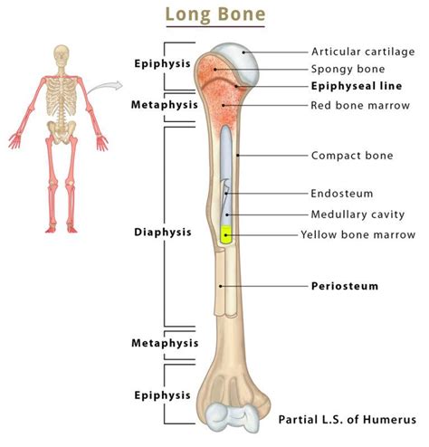
Table of Contents
Labeling the Structures of a Long Bone: A Comprehensive Guide
Long bones, the foundational elements of our limbs, are fascinating structures with intricate designs perfectly tailored for their weight-bearing and movement functions. Understanding their anatomy is crucial for anyone studying biology, medicine, or related fields. This comprehensive guide will walk you through the process of labeling the various structures of a long bone, providing detailed explanations and high-yield insights to aid your learning.
The Key Components of a Long Bone
Before diving into labeling, let's familiarize ourselves with the major structures found in a typical long bone. These structures work in concert to provide strength, support, and facilitate growth and repair. Remember that the specific dimensions and features might slightly vary depending on the individual bone and location in the body.
1. Diaphysis (Shaft): The Central Core
The diaphysis, or shaft, forms the long, cylindrical main portion of the long bone. It's primarily composed of compact bone, a dense and strong type of bone tissue offering significant resistance to stress and strain. This dense structure is essential for weight-bearing and protecting the internal structures. The diaphysis's exterior is covered by a fibrous membrane called the periosteum, which plays a critical role in bone growth, repair, and nutrient supply.
2. Epiphyses: The Growth Plates and Articulating Ends
At either end of the diaphysis lie the epiphyses, the wider, more bulbous portions of the bone. These are primarily composed of spongy bone, also known as cancellous bone. Spongy bone has a porous, honeycombed structure, making it lighter than compact bone while still providing substantial strength. The spaces within the spongy bone contain red bone marrow, responsible for the production of red blood cells, white blood cells, and platelets – a vital process known as hematopoiesis.
The epiphyses are covered with a layer of articular cartilage, a smooth, resilient tissue that reduces friction and absorbs shock at the joints. This smooth surface allows for easy articulation and movement with other bones.
3. Metaphyses: The Transition Zones
Between the diaphysis and epiphyses are the metaphyses. These transitional zones are crucial during bone growth. In children and adolescents, the metaphyses contain the epiphyseal plates, also known as growth plates. These are cartilaginous regions where bone lengthening occurs. Once growth is complete, the epiphyseal plates ossify (turn into bone), forming the epiphyseal lines.
4. Medullary Cavity: The Inner Space
Within the diaphysis lies the medullary cavity, a hollow space filled with yellow bone marrow in adults. Unlike red bone marrow, yellow bone marrow primarily consists of fat cells and plays a less significant role in blood cell production. However, in times of severe blood loss, yellow bone marrow can revert to red bone marrow to increase blood cell production.
5. Periosteum and Endosteum: The Protective Layers
The periosteum, as mentioned earlier, is a fibrous membrane covering the outer surface of the bone except for the articular cartilage at the joints. It's richly supplied with blood vessels and nerves and plays a vital role in bone growth, repair, and nutrient supply. The periosteum also serves as an attachment point for tendons and ligaments.
The endosteum is a thin membrane lining the inner surface of the bone, including the medullary cavity. It contains bone-forming cells (osteoblasts) and bone-resorbing cells (osteoclasts), contributing to bone remodeling and maintenance.
6. Nutrient Foramina: The Supply Lines
Running through the periosteum and into the bone are numerous small openings called nutrient foramina. These foramina provide pathways for blood vessels and nerves to enter the bone, supplying it with essential nutrients and oxygen and allowing for sensory feedback.
Step-by-Step Guide to Labeling a Long Bone Diagram
Now, let’s put this knowledge into practice. You will need a diagram of a long bone (a femur or humerus works well). Follow these steps:
-
Identify the Diaphysis: Locate the long, cylindrical shaft of the bone. This is the diaphysis. Label it clearly.
-
Locate the Epiphyses: Identify the broader ends of the bone. These are the epiphyses. Label the proximal epiphysis (the end closer to the body's center) and the distal epiphysis (the end farther from the body's center).
-
Mark the Metaphyses: Find the areas where the diaphysis meets the epiphyses. These are the metaphyses. Label them accordingly. If dealing with a young bone, look for the epiphyseal plates within the metaphyses. In adult bones, you'll find the epiphyseal lines instead. Label these as well.
-
Indicate the Medullary Cavity: Locate the hollow space within the diaphysis. This is the medullary cavity. Label it clearly. Specify whether it contains yellow bone marrow (in adults) or predominantly red bone marrow (in infants and children, although red marrow is also found in specific areas in adult bones).
-
Highlight the Periosteum: Indicate the fibrous membrane covering the external surface of the bone. This is the periosteum.
-
Show the Endosteum: Locate the thin membrane lining the inner surface of the bone, especially within the medullary cavity. This is the endosteum.
-
Point out the Articular Cartilage: Identify the smooth cartilage covering the articular surfaces of the epiphyses. Label it accordingly. This is crucial for smooth joint movement.
-
Identify Nutrient Foramina: Look for small openings on the surface of the bone. These are the nutrient foramina. Label a few of them to show their distribution.
-
Differentiate Compact and Spongy Bone: Within the diaphysis, the dense outer layer is compact bone. In the epiphyses, the more porous inner layer is spongy bone. Indicate these regions on your diagram.
Clinical Significance of Understanding Long Bone Structure
Understanding the structure of long bones is critical for diagnosing and treating various musculoskeletal conditions. Here are some examples:
-
Fractures: Knowing the different parts of a long bone helps clinicians determine the type and severity of a fracture, influencing the treatment strategy. For example, a fracture involving the epiphyseal plate in a child can have long-term implications for bone growth.
-
Osteoporosis: Understanding bone structure helps diagnose and monitor osteoporosis, a condition characterized by decreased bone density, which increases the risk of fractures, particularly in the long bones.
-
Bone Infections (Osteomyelitis): Knowledge of the bone's structure helps clinicians understand how infections can spread within the bone, impacting treatment decisions, such as surgical debridement or antibiotic administration.
-
Bone Tumors: The location of a bone tumor within the long bone, whether in the diaphysis, metaphysis, or epiphysis, affects treatment planning, surgical approaches, and prognosis.
-
Bone Marrow Transplantation: Understanding bone marrow location is vital for bone marrow transplantation procedures. The specific areas for aspiration or injection are determined by knowledge of the medullary cavity's location and the distribution of red and yellow bone marrow.
Beyond the Basics: Microscopic Structure and Cellular Components
While this guide focuses on the macroscopic structure of long bones, it's important to remember that understanding the microscopic anatomy is equally important. This includes:
-
Osteocytes: Mature bone cells responsible for maintaining bone tissue.
-
Osteoblasts: Bone-forming cells that synthesize and deposit new bone matrix.
-
Osteoclasts: Bone-resorbing cells that break down bone tissue, crucial for bone remodeling and calcium homeostasis.
-
Haversian Systems (Osteons): The basic structural units of compact bone, consisting of concentric lamellae surrounding a central canal containing blood vessels and nerves.
-
Lacunae and Canaliculi: Small spaces within the bone matrix where osteocytes reside, connected by tiny canals called canaliculi allowing for nutrient and waste exchange.
Understanding these cellular components and their functions is crucial for comprehending the dynamic nature of bone tissue and its ability to remodel and repair itself throughout life.
Conclusion
Labeling the structures of a long bone is a fundamental step in mastering skeletal anatomy. This comprehensive guide, encompassing both macroscopic and microscopic perspectives, equips you with the knowledge to accurately identify and understand the functions of each component. Remember to practice labeling diagrams regularly to solidify your understanding. This detailed knowledge is not only essential for academic pursuits but also forms the cornerstone for understanding various clinical conditions related to the skeletal system. By understanding the intricacies of long bone structure, you gain a deeper appreciation for the remarkable engineering of the human body.
Latest Posts
Latest Posts
-
Which Of The Following Is A Complete Sentence
Mar 21, 2025
-
Name The Three Main Types Of Intaglio Printing
Mar 21, 2025
-
What Is Unique About An Azeotropic Refrigerant Mixture
Mar 21, 2025
-
The Region Of High Hydrogen Ion Concentration Is The
Mar 21, 2025
-
The 2024 Final Rule Specifically Defines What Qualifies As Consent
Mar 21, 2025
Related Post
Thank you for visiting our website which covers about Label The Structures Of A Long Bone . We hope the information provided has been useful to you. Feel free to contact us if you have any questions or need further assistance. See you next time and don't miss to bookmark.
