Optic Nerve And Blood Vessels Enter The Eye At The
Breaking News Today
Apr 04, 2025 · 7 min read

Table of Contents
Optic Nerve and Blood Vessels Enter the Eye at the Optic Disc: A Comprehensive Overview
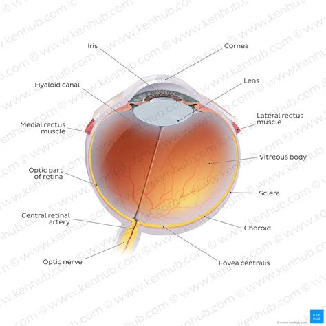
The human eye, a marvel of biological engineering, relies on a complex interplay of structures to function effectively. Central to this intricate system is the optic disc, the point where the optic nerve and retinal blood vessels enter and exit the eye. Understanding the anatomy, physiology, and potential pathologies associated with this critical area is crucial for comprehending ocular health and disease. This article will delve into the detailed structure and function of the optic disc, exploring its significance in vision and highlighting common associated conditions.
Anatomy of the Optic Disc: A Detailed Look
The optic disc, also known as the blind spot, is a roughly circular region located on the nasal side of the retina. Its size typically ranges from 1.5 to 2 millimeters in diameter. This seemingly insignificant area plays a pivotal role in visual perception and overall eye health. Several key anatomical features define the optic disc:
1. Optic Nerve Head: The Exit Point
The optic nerve head is the most prominent feature of the optic disc. This is the point where the axons of retinal ganglion cells converge to form the optic nerve (CN II), exiting the eyeball to transmit visual information to the brain. The nerve fibers are meticulously organized as they exit the eye, forming a distinct structure visible during ophthalmoscopic examination. The appearance of the optic nerve head, including its size, shape, and color, can provide valuable diagnostic clues about underlying eye conditions.
2. Retinal Blood Vessels: Supply and Drainage
The optic disc serves as the entry point for the central retinal artery and its branches, supplying oxygen and nutrients to the retina. These vessels are readily visible during ophthalmoscopic examination, branching out from the optic disc to irrigate the entire retinal surface. Similarly, the central retinal vein and its branches exit the eye at the optic disc, draining metabolic waste products from the retina. The arrangement and caliber of these blood vessels are critical indicators of retinal health and can reveal evidence of vascular diseases.
3. Peripapillary Retina: Surrounding Tissue
The area surrounding the optic disc, known as the peripapillary retina, is a region of unique anatomical and functional significance. This region lacks photoreceptor cells, contributing to the blind spot in our visual field. However, its neural composition and proximity to the optic nerve head make it a critical area for assessing retinal health and identifying early signs of neurological diseases. The peripapillary retina often exhibits distinct structural features that aid in diagnosing retinal pathologies.
4. Lamina Cribrosa: A Sieve-like Structure
The lamina cribrosa is a sieve-like structure composed of collagenous and elastic fibers that forms the interface between the sclera and the optic nerve head. This structure acts as a supportive framework for the optic nerve fibers as they exit the eye. Its structural integrity is crucial for maintaining the structural integrity of the optic nerve head. Changes in the lamina cribrosa, such as thinning or deformation, can be a hallmark of glaucoma.
Physiology of the Optic Disc: Vision and Beyond
The optic disc's physiological role extends beyond being a mere entry/exit point for the optic nerve and blood vessels. Its intricate structure and location influence various visual processes:
1. Visual Signal Transmission
The primary function of the optic disc is to facilitate the transmission of visual signals from the retina to the brain. The retinal ganglion cell axons, carrying encoded visual information, converge at the optic disc, forming the optic nerve. This nerve then transmits the signals to the lateral geniculate nucleus (LGN) in the thalamus and eventually to the visual cortex, where the visual signals are interpreted. Any disruption to this pathway, often caused by damage to the optic nerve head, can lead to vision loss.
2. Blood Supply to Retina
The central retinal artery, entering the eye at the optic disc, provides the retina with its vital blood supply. This artery branches into smaller arterioles and capillaries, ensuring efficient oxygen and nutrient delivery to the retinal cells. Compromised blood flow, due to conditions like retinal artery occlusion, can cause irreversible retinal damage and vision loss. The venous system, draining metabolic waste, is equally important in maintaining retinal homeostasis.
3. Maintenance of Intraocular Pressure
The optic disc’s structural integrity plays a crucial role in maintaining the intraocular pressure (IOP) of the eye. The lamina cribrosa's ability to withstand IOP fluctuations is vital in preventing damage to the optic nerve head. Elevated IOP, a hallmark of glaucoma, can lead to deformation of the lamina cribrosa and subsequent optic nerve damage, resulting in characteristic visual field loss.
Clinical Significance of the Optic Disc: Common Conditions
The optic disc's importance is underscored by its involvement in numerous ocular pathologies. Observing and assessing the appearance of the optic disc during ophthalmoscopic examination is a critical component of routine eye exams.
1. Glaucoma: Damage to Optic Nerve Head
Glaucoma, a group of eye diseases characterized by elevated IOP and progressive optic nerve damage, often presents with characteristic changes in the optic disc. These changes include cupping of the optic disc (increased depth of the central depression), thinning of the neuroretinal rim, and the appearance of nerve fiber layer defects. Early detection of glaucoma is critical to preventing irreversible vision loss, emphasizing the significance of regular eye exams.
2. Papilledema: Swelling of the Optic Disc
Papilledema, characterized by swelling of the optic disc, is often a sign of increased intracranial pressure (ICP). The swelling results from impaired venous drainage from the optic nerve head. Papilledema necessitates a thorough neurological evaluation to identify the underlying cause, which may include brain tumors, meningitis, or other serious neurological conditions.
3. Optic Neuritis: Inflammation of Optic Nerve
Optic neuritis, an inflammatory condition affecting the optic nerve, can lead to swelling and discoloration of the optic disc. This condition is often associated with multiple sclerosis and other autoimmune diseases. Prompt diagnosis and treatment of optic neuritis are critical to minimizing vision loss and preventing long-term complications.
4. Ischemic Optic Neuropathy: Reduced Blood Flow
Ischemic optic neuropathy, resulting from reduced blood flow to the optic nerve head, can lead to sudden vision loss. This condition can be caused by a variety of factors, including vascular diseases and systemic conditions. Prompt diagnosis and management are crucial to minimizing vision loss and preventing complications.
5. Optic Disc Drusen: Deposits in the Optic Nerve Head
Optic disc drusen are hyaline deposits that accumulate in the optic nerve head. While often asymptomatic, they can sometimes cause visual disturbances and mimic other optic disc pathologies. Understanding the presence of optic disc drusen is important in differentiating them from other potentially sight-threatening conditions.
Diagnostic Tools for Evaluating the Optic Disc
Several diagnostic tools are used to evaluate the optic disc and assess its health:
- Ophthalmoscopy: Direct and indirect ophthalmoscopy provides a detailed view of the optic disc, allowing for assessment of its color, size, shape, and the condition of its surrounding structures, including the retinal blood vessels.
- Optical Coherence Tomography (OCT): OCT provides high-resolution cross-sectional images of the optic nerve head and retinal layers, enabling precise measurements of the retinal nerve fiber layer thickness and the depth of optic disc cupping.
- Visual Field Testing: Visual field testing assesses the extent of vision loss, identifying areas of visual field defects associated with optic nerve damage. This is particularly useful in diagnosing glaucoma.
- Fluorescein Angiography: Fluorescein angiography is an imaging technique that visualizes the retinal vasculature, allowing for assessment of blood flow and the detection of vascular abnormalities. This technique can be crucial in diagnosing vascular diseases impacting the optic disc.
Conclusion: The Optic Disc as a Window to Ocular and Neurological Health
The optic disc, while seemingly a small and inconspicuous area, is a critical structure with significant clinical importance. Its role as the entry and exit point for the optic nerve and retinal blood vessels makes it a valuable indicator of both ocular and neurological health. Understanding the anatomy, physiology, and common pathologies associated with the optic disc is vital for ophthalmologists and other healthcare professionals in diagnosing and managing a wide range of eye and neurological conditions. Regular eye examinations, including a comprehensive evaluation of the optic disc, are essential for early detection and timely intervention, ultimately preserving vision and overall health. The continuing advancements in diagnostic imaging and treatment strategies ensure that our understanding of the optic disc and its significance continues to evolve, leading to improved patient care.
Latest Posts
Latest Posts
-
Model 1 Movement Of Water In And Out Of Cells
Apr 04, 2025
-
Why Do Cell Phones Present A Problem For Pollsters
Apr 04, 2025
-
Which Of The Following Is True Of Human Resource Planning
Apr 04, 2025
-
Points Of Distribution Are Strategically Placed Facilities Where
Apr 04, 2025
-
The Meeting Of People As A Romantic Engagement
Apr 04, 2025
Related Post
Thank you for visiting our website which covers about Optic Nerve And Blood Vessels Enter The Eye At The . We hope the information provided has been useful to you. Feel free to contact us if you have any questions or need further assistance. See you next time and don't miss to bookmark.
